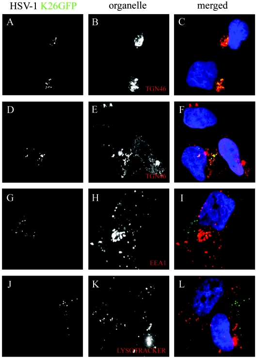
FIG. 5.
Colocalization of capsids with the TGN. 143B cells were infected with wild-type K26GFP virus (in green), incubated for 7 h at 37°C, and chased at 20°C for 6 h. They were then fixed and stained for various subcellular markers (in red), including TGN46 (A to F), EEA1 (G to I) and Lysotracker (J to L).

