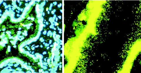
FIG. 2.
Residual virions from the inoculum trapped in cervical mucus. Sections of a cervix obtained 24 h after intravaginal exposure to SIV were cut and hybridized to SIV-specific riboprobes, and the SIV RNA signal from virions or infected cells was amplified by TSA/ELF as described in Materials and Methods. Viral RNA was not detected in cells but was detected in virions, as shown in the mucus adhering to the epithelium of the endocervical gland shown. (Left) Original magnification, ×100. The image brightness and contrast were adjusted to reveal Hoechst-counterstained nuclei and histology. (Right) Magnification, ×1,000. The image was adjusted to reveal virions at the mucosal surface.

