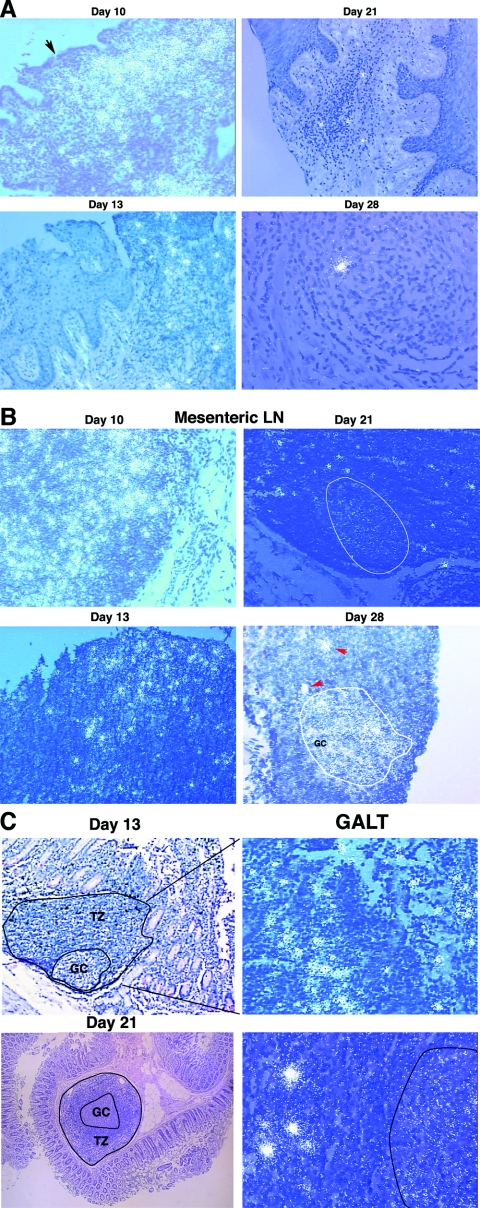
FIG. 6.
Examples of SIV RNA-positive cells in cervicovaginal and lymphatic tissues and of virions associated with follicular dendritic cells (FDCs) in LTs. SIV RNA in cells and virions was detected by ISH with 35S-labeled riboprobes. In the developed radioautographs illuminated with epipolarized light, SIV RNA-positive cells appear as bright ob-jects and FDC-associated virions appear as bright silver grains overlying follicles. (A) Cervical and vaginal tissues. On day 10, there were dense collections of SIV RNA-positive cells in inflammatory foci in the submucosa of the endocervix. The arrow points to a break in the overlying epithelium. SIV RNA-positive cells progressively decreased in frequency from days 13 to 28 and were usually associated with small foci of inflammation. (B) LNs. In mesenteric and other LNs, there were numerous SIV RNA-positive cells on day 10. Their numbers then declined. A faint signal from FDC-associated virion RNA in follicles (encircled) was detectable by day 21 and increased by day 28 in the encircled germinal center of a follicle. The red arrows each point to SIV RNA-positive cells in the adjacent T-cell zone. (C) GALT. On day 13, dense collections of SIV RNA-positive cells in the follicular aggregate of this section from the jejunum were evident in the T-cell zone (TZ; enlarged view, right panel). On day 21, FDC-associated virion RNA was detectable in the outlined germinal center (GC), in addition to SIV RNA-positive cells (right panel).

