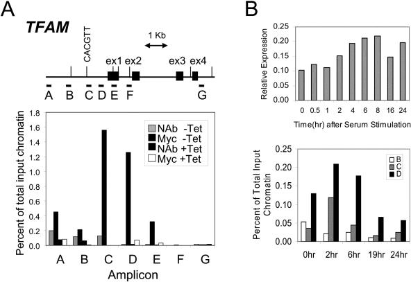
FIG. 6.
c-Myc binds to TFAM in situ. (A) Human genomic sequence starting from 3 kb upstream of exon 1 to 7 kb downstream. Exons are represented by black boxes. The E boxes are indicated with vertical bars, and the E box in amplicon C is illustrated. Horizontal bars labeled A to G indicate the regions amplified for scanning ChIP analysis. P493-6 cells were plated in absence (−Tet) or presence (+Tet) of tetracycline as described in Materials and Methods, and then ChIP was performed with anti-c-Myc antibody (Myc −Tet or Myc +Tet). No-antibody control experiments (NAb −Tet or NAb +Tet) were performed at same time. −Tet corresponds to a high-Myc state, whereas +Tet represents a low-Myc state. Quantitative PCRs were performed. Shown are averages of triplicates. (B) Top, time-dependent expression of TFAM following serum stimulation of human 2091 primary fibroblasts. TFAM expression is shown as normalized expression relative to 18S rRNA. Mean values (with standard deviations of less than 5% of the mean) from triplicate real-time PCRs are shown. Bottom, chromatin immunoprecipitation assays showing time-dependent binding of Myc to TFAM following serum stimulation of human 2091 primary fibroblasts. Binding to amplicons B, C, and D, which are defined in panel A, is shown as a percentage of total input DNA.

