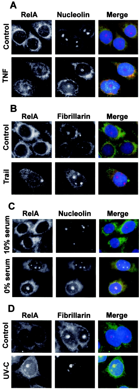
FIG.3.
Specificity of stimuli causing nucleolar localization of RelA. (A) SW480 cells were unstimulated (control) or stimulated with TNF (10 ng/ml, 1 h) and then fixed, and immunocytochemistry was performed using specified antibodies. Nucleoli are stained with nucleolin. (B) Immunocytochemistry was performed on SW480 cells either untreated or treated with TRAIL (10 ng/ml) for 16 h. Fibrillarin was used to identify nucleoli. (C) SW480 cells were seeded in medium containing 10% serum and grown for 16 h. Medium was then replaced by fresh medium containing either 10% or 0% serum, and cells were grown for a further 96 h. Following fixation, immunocytochemistry was performed using antibodies to RelA and nucleolin as described above. (D) Immunocytochemistry, using anti-RelA and anti-nucleolin antibodies, was performed on SW480 cells 5 h after mock or UV-C (40 J/m2) irradiation. In all merged panels, DNA is stained by DAPI and appears blue.

