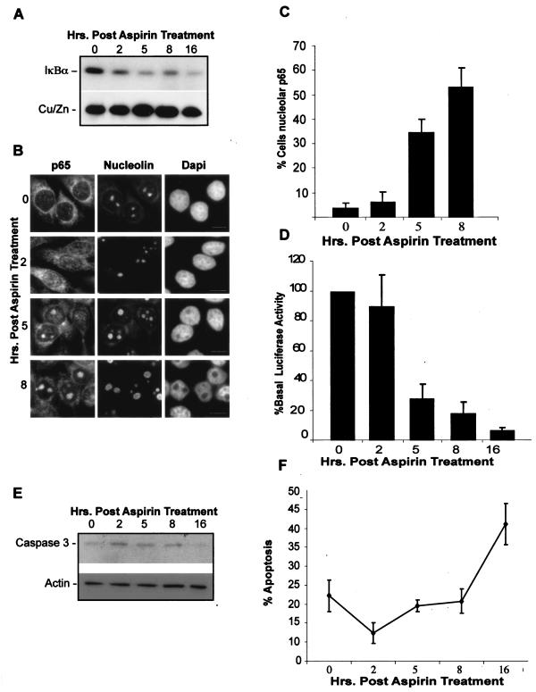
FIG. 7.
IκB degradation, nucleolar translocation of RelA, repression of NF-κB transcription, and apoptosis occur sequentially. (A) IκB degradation occurs 2 to 5 h after stimulation with aspirin. Cells were treated with aspirin (10 mM) for 0 to 16 h, and then cytoplasmic IκBα and control protein (CU/Zn SOD) levels were determined using Western blot analysis. (B and C) Nucleolar accumulation of RelA (p65) is apparent 5 to 8 h after aspirin stimulation. Immunocytochemistry was performed on SW480 cells treated as described above. DAPI staining depicts nuclei. Nucleoli are stained with nucleolin. Bars, 10 μm. The percentage of cells in the population showing nucleolar RelA (p65) was quantified. Data are the means of at least three experiments (± SE). (D) Decreased NF-κB-driven transcription occurs concurrently with nucleolar accumulation of RelA. SW480 cells were transfected with the NF-κB dependent 3xκB ConA-luc and the control pCMVβ reporter constructs. Twenty-four hours after transfection, cells were treated as described above. Values are presented as the percentages of luciferase activity at time zero and are the means (± SE) of three independent experiments after β-galactosidase normalization. (E) Aspirin-induced caspase-3 cleavage occurs 16 h after stimulation. SW480 cells were treated as described above and levels of uncleaved caspase-3 determined by Western blot analysis performed on cytoplasmic extracts. (F) Percentages of SW480 cells undergoing apoptosis, as determined by annexin V staining, following aspirin treatment as described above.

