FIG. 2.
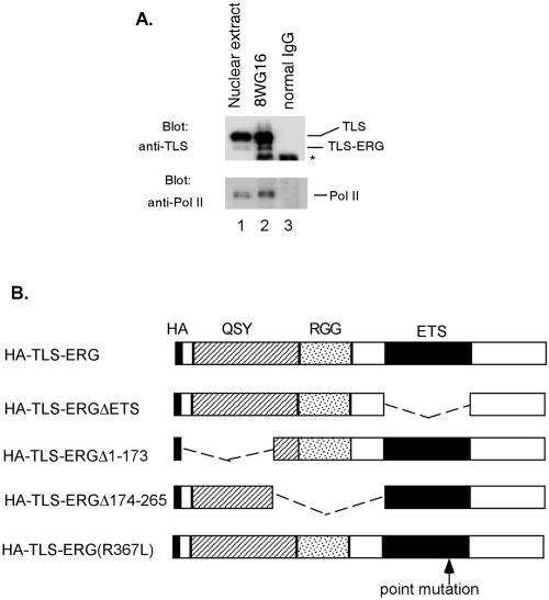
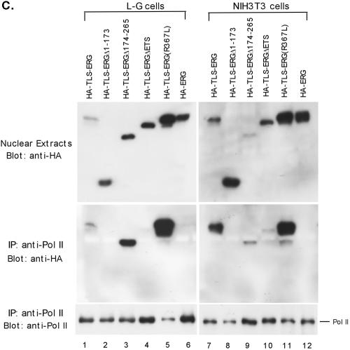
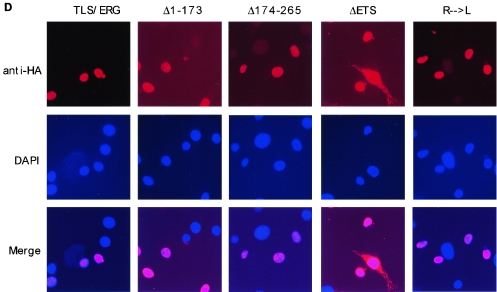
Interaction of RNA Pol II with the first 173 amino acids of TLS-ERG. (A) YNH-1 nuclear extract (lane 1) was incubated with the anti-Pol II antibody 8WG16 (lane 2) or normal mouse IgG as a negative control (lane 3). The position of the IgG band is indicated by an asterisk. (B) Schematic of HA-TLS-ERG and its mutants with different domains deleted or mutated. HA, hemagglutinin epitope; QSY, glutamine-, serine-, and tyrosine-rich domain; RGG, region with multiple Arg-Gly-Gly repeats; ETS, ets DNA-binding domain. (C) Nuclear extracts from L-G cells (lanes 1 to 6) and NIH 3T3 cells (lanes 7 to 12) expressing HA-TLS-ERG or its mutants were blotted with the 3F10 anti-HA antibody (top panels). The nuclear extracts were incubated with 8WG16, and the immunoprecipitates (IP) were blotted with the anti-HA antibody (middle panels) or the C21 anti-Pol II antibody (bottom panels). (D) NIH 3T3 cells expressing HA-TLS-ERG or its mutants were stained with a Cy3-conjugated anti-HA antibody (top panel), the nuclei were indicated by DAPI staining (middle panel), and the two images were merged to show subcellular localization of the HA-tagged protein.


