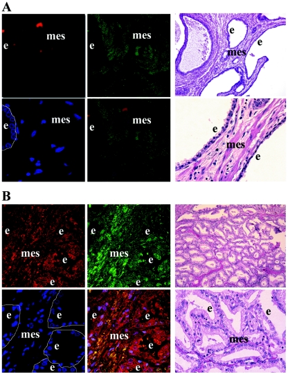
FIG. 8.
Localization of PDGF D and uPA in human prostate carcinoma. Microscopic immunofluorescence analysis of uPA (Texas Red) and PDGF D (fluorescein isothiocyanate; green) expression in samples of the human prostate was performed at 63× magnification. Cell nuclei were stained with 4′,6′-diamidino-2-phenylindole (DAPI; blue). Yellow in the merged panel represents the colocalization of uPA and PDGF D. Images of H&E staining of corresponding serial slides taken at 10× and 40× magnification were used for histological analysis of tissues. Epithelial cells lining the ducts of the prostate are outlined in the DAPI panels. e, epithelium; mes, mesenchyme. (A) Normal human prostate specimen. (B) Human prostate carcinoma specimen.

