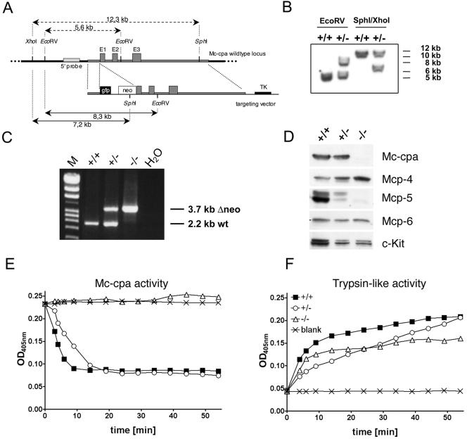
FIG. 1.
Generation of Mc-cpa−/− mice and protein and enzyme analyses of Mc-cpa−/− mast cells. (A) The targeting construct included a short 5′ arm, the enhanced green fluorescent protein gene (gfp), the neomycin resistance gene (neo), a long 3′ arm including exons 1 (from nucleotide +18 on), 2 and 3, and the thymidine kinase (TK) gene. (B) Southern blot of Mc-cpa+/+ (+/+) and Mc-cpa+/− (+/−) ES cell DNA digested with EcoRV or SphI/XhoI. The 5′ probe and expected fragment lengths are shown in panel A. (C) Mc-cpa+/+, Mc-cpa+/−, and Mc-cpa−/− mice were identified by single 2.2-kb PCR bands, 2.2-kb and 3.7-kb PCR bands, or single 3.7-kb PCR bands. Lane M contains molecular size markers. wt, wild type. (D) Western blot analysis of Mc-cpa+/+, Mc-cpa+/−, and Mc-cpa−/− mast cells shows the absence of Mc-cpa and Mcp-5 proteins in Mc-cpa−/− mast cells. Mcp-4 is upregulated, and Mcp-6 is unaltered. (E and F) Lysates of Mc-cpa+/+ (▪), Mc-cpa+/− (○), and Mc-cpa−/− (▵) peritoneal mast cells or the blank control (×) were analyzed for carboxypeptidase activity (E) and trypsin-like activity (F) by test substrates (15). Mc-cpa−/− mast cells lacked Mc-cpa activity (E). Cells from all genotypes had trypsin-like activity (F). OD405nm, optical density at 405 nm.

