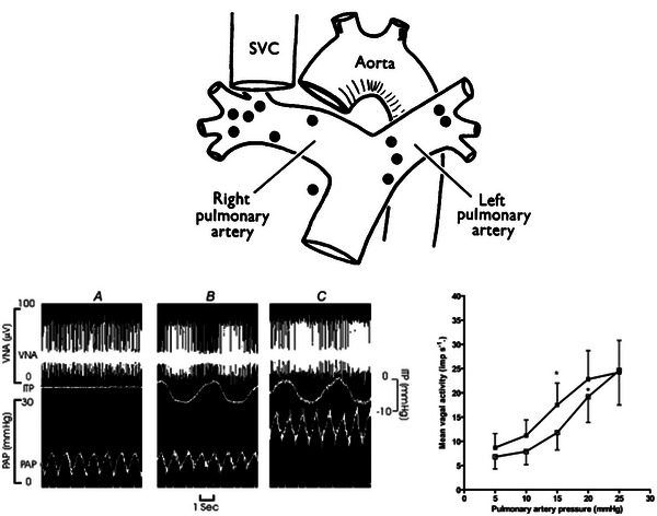
FIGURE 3.

The top panel shows the location of vagal afferent pulmonary arterial baroreceptors as determined by electrophysiological testing in dogs. Receptors were located predominately in the main pulmonary artery, its bifurcation and the proximal right and left pulmonary artery branches. Reproduced with permission from Coleridge & Kidd (1960). Abbreviation: SVC, superior vena cava. The bottom panels depict pulmonary baroreceptor vagal nerve activity (VNA) in close‐chested dogs with a vascularly isolated pulmonary circulation, reproduced with permission from Moore et al. (2004b). In the left panel, an original pulmonary baroreceptor VNA recording is shown from one dog, with pulsations of pulmonary artery pressure (PAP) without negative phasic intrathoracic pressure (ITP; A), VNA with negative phasic ITP (B) and increased VNA with a step increase in PAP with negative phasic ITP (C). The right panel shows the mean VNA with increases in PAP at non‐phasic atmospheric ITP (open squares) and negative phasic ITP (filled squares). The mean threshold PAP for VNA response reduced from 12 to 9.5 mmHg with negative phasic ITP. * P < 0.05 compared with baseline VNA.
