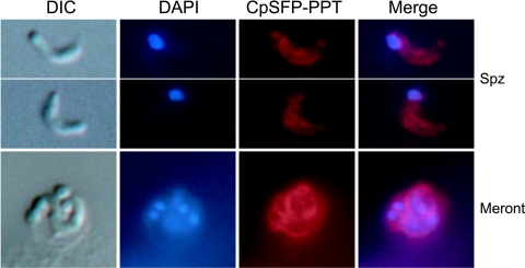
FIG. 6.
Indirect immunofluorescence microscopic detection of CpSFP-PPT in C. parvum free sporozoites (Spz) and an intracellular developmental stage (a merozoite-containing meront) cultured in HCT-8 cells for 24 h. Samples were labeled with rabbit polyclonal antibody to CpSFP-PPT and TRITC-conjugated antirabbit immunoglobulin G monoclonal antibody that gave a diffused pattern in the cytosol of all samples. Preimmune serum did not label the parasite (data not shown). The nuclei were counterstained with 4′,6′-diamidino-2-phenylindole (DAPI). Parasite morphology is shown as differential interference constrast (DIC) images taken from the same microscopic fields. A similar pattern of cytosolic distribution of CpSFP-PPT was observed in other intracellular stages (e.g., 12 to 72 h postinfection) but is not shown here.

