
FIG. 1.
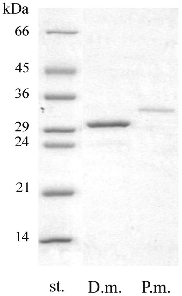
SDS-PAGE of the purified keratinases of P. marquandii (P.m.) and D. microsporus (D.m.). The positions of low-molecular-mass markers (st.) from Sigma are shown. The gel (12%) was stained with Coomassie brilliant blue.
Official websites use .gov
A
.gov website belongs to an official
government organization in the United States.
Secure .gov websites use HTTPS
A lock (
) or https:// means you've safely
connected to the .gov website. Share sensitive
information only on official, secure websites.
SDS-PAGE of the purified keratinases of P. marquandii (P.m.) and D. microsporus (D.m.). The positions of low-molecular-mass markers (st.) from Sigma are shown. The gel (12%) was stained with Coomassie brilliant blue.