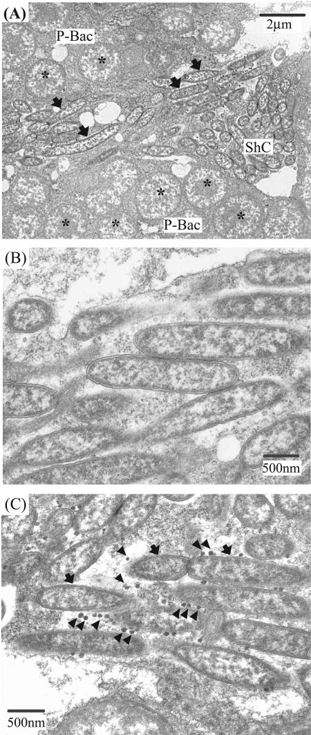
FIG. 3.
Electron microscopy of the Rickettsia symbiont. (A) A sheath cell harboring rod-shaped cells of the Rickettsia symbiont (arrows) surrounded by primary mycetocytes harboring Buchnera (asterisks). (B) A magnified image of the Rickettsia cells in a sheath cell. (C) Virus-like particles (arrowheads) in association with the Rickettsia cells. Abbreviations: ShC, sheath cell; PM, primary mycetocyte.

