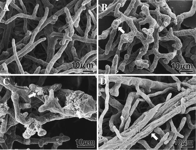
FIG. 2.
Scanning electron micrographs of A. cochlioides mycelial samples interacting (5 days) with Lysobacter sp. strain SB-K88 and an untreated control. For the SEM study, small blocks (diameter, 6 mm) of affected mycelia (approaching a bacterial colony) in agar were transferred from a petri dish (inside diameter, 9 cm) containing corn meal agar to a 3-cm-inside-diameter petri dish on day 6 of cultivation and then fixed with 2% glutaraldehyde in phosphate buffer (8 mM, pH 7.2) for 3 h. Other preparation procedures for microscopy were similar to those described previously (14, 17, 18). (A) Normal growth in the absence of SB-K88 (control). (B) Bulbous structures (arrow) and curly growth in the presence of SB-K88. (C) Cytoplasmic extrusion from the hyphae (arrows). (D) Overlapping growth of mycelia (arrow).

