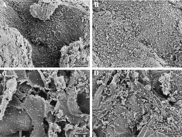
FIG. 3.
Scanning electron micrographs showing seed coats of spinach (A and B) and sugar beet (C and D) at zero time (A and C) and 48 h (B and D) after inoculation of seeds with Lysobacter sp. strain SB-K88 cells (see Materials and Methods for details). The numbers of bacteria per 100 μm2 of seed coat determined by SEM were 69 ± 8 cells (zero time) and 158 ± 11 cells (48 h) for spinach and 22 ± 4 cells (zero time) and 53 ± 9 cells (48 h) for sugar beet. These values are averages ± standard errors of five replications.

