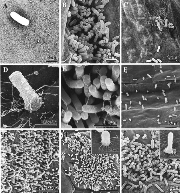
FIG. 4.
TEM (A) and SEM (B to I) micrographs illustrating the morphology of Lysobacter sp. strain SB-K88 (A) and colonization of SB-K88 (B to I) on plant surfaces upon inoculation of seeds and seedlings grown in the gellan gum-based medium (B to D and G to I) or soil (F). (A) TEM micrograph of a sessile SB-K88 bacterial cell having large, brush-like fimbriae at one end. (B) Colonization on sugar beet root by perpendicular attachment. (C) Bacterial biofilm that developed under a semitransparent film of sugar beet root mucigel. (D) Typical perpendicular attachment of a bacterial cell to a sugar beet cotyledon. (E) High-density perpendicular attachment and colonization on the sugar beet leaf surface after immersion into an SB-K88 bacterial suspension (ca. 105 CFU/ml). (F) Colonization of sugar beet root. (G) Colonization of tomato root. (H) Colonization of A. thaliana leaf. (I) Colonization of A. thaliana root. Each experiment was repeated three times, and representative micrographs are shown (see Materials and Methods for details).

