FIG. 6.
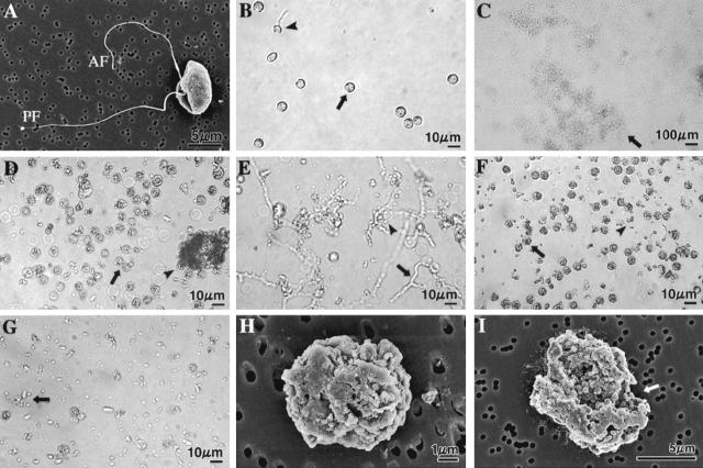
Light (B to G) and SEM (H and I) micrographs showing A. cochlioides zoospore-lytic activity of the freeze-dried culture supernatant, EtOAc- and water-soluble fractions of the culture supernatant, and pure xanthobaccin A. The micrographs in panels B to G were taken after 3 h of treatment by focusing on the bottom of a petri dish with a digital camera connected to the microscope (for details see Materials and Methods). Crude extracts or pure xanthobaccin A (dissolved in small quantities of DMSO) at the concentrations tested immediately caused inhibition of the motility of zoospores (see Table 1 and Materials and Method for details of the bioassay method). The halted zoospores rapidly settled to the bottom of the dish and then started to burst or lyse. The final concentration of DMSO in the aqueous zoospore suspension was maintained at less than 1% in all treatments. DMSO alone (final concentration, 1%) was used as the negative control and caused no lysis of zoospores. Each experiment was replicated at least five times, and representative micrographs are shown. (A) SEM micrograph of a biflagellate A. cochlioides zoospore (untreated control). AF, anterior flagellum; PF, posterior flagellum. (B) No lysis in the control dish (1% DMSO). A small portion (10 to 15%) of the motile zoospores in the control dish were stopped and changed into round cystospores (arrow) after 3 h and then settled to the bottom of the dish; 5 to 8% of these cystospores germinated (arrowhead) and formed germ tubes. No motile zoospores were observed in aqueous medium because the photograph was taken by focusing on bottom of the dish. (C) Complete lysis of all halted zoospores by freeze-dried culture supernatant (500 μg/ml). The arrow indicates lysed material. (D) All spores were granulated or lysed (arrow) by the EtOAc-soluble fraction (100 μg/ml). Some lysed material aggregated (arrowhead). (E) Water-soluble fraction (500 μg/ml) initially induced germination of cystospores (arrow and arrowhead) within 1 h, and then (3 h) all spores and germ tubes were partially lysed (arrow and arrowhead). (F) Xanthobaccin A (1 μg/ml) caused granulation (arrowhead) and lysis (arrowhead) of all spores. (G) Complete lysis of zoospores (arrow) by xanthobaccin A (1 μg/ml). (H and I) Scanning electron micrographs of granulated, cracked, and lysed (arrow in I) A. cochlioides zoospores exposed to 1 ppm xanthobaccin A for 30 min.

