FIG. 7.
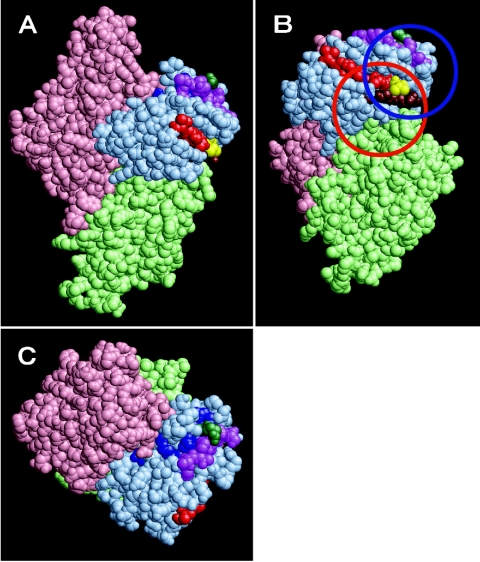
Epitopes of blocking antibodies in a three-dimensional model of Cry1Aa. (A) The amino acid residues in domains I, II, and III are pink, light green, and light blue, respectively. The single octapeptide to which MAb 1B10 bound, 582VFTLSAHV589, is red. The three octapeptides to which MAb 2C2 bound, 520QRYRVRIR527, 570FTTPFNFS577, and 582VFTLSAHV589 are blue, magenta, and red, respectively. In addition, Arg521 (green) and Val582 (yellow), each of which was mutated to cysteine, are shown. The peptide 508STLRVN513 (brown) adjoins the 582VFTLSAHV589 site in the three-dimensional structure; this region is expected to be involved in the binding of the blocking MAbs. The candidate binding sites for MAbs 1B10 and 2C2 are indicated by blue and red circles. (B) Panel A rotated 72° about the y axis and 45° about the x axis. (C) Panel A rotated 90° about the x axis.

