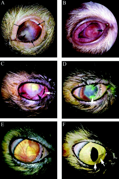
FIG. 1.
(A) Case 1. Corneal herpetic ulcer and stromal ulceration in the left eye of a 15-year-old Birman cat at presentation. The thin long black arrows delineate the edge of the large ulceration, which is surrounded by corneal vessels. The short black arrow points to the center of the stromal area of corneal necrosis. (B) Case 2. Severe corneal necrosis of a 7-week-old domestic shorthair cat at presentation. The short black arrow indicates the cream-colored infiltrate and corneal necrosis. The entire cornea was vascularized. Raised corneal vascularization is present to the left of the necrotic lesion. (C) Case 3. Severe corneal ulceration in the right eye of a 1.5-year-old domestic shorthair cat at presentation. The conjunctiva is moderately hyperemic, and the nictitating membrane (white arrow) is prolapsed. The cornea is vascularized from the limbus to the stromal ulcer (black arrows). The cream-colored necrotic margins of the ulcer can be seen at the black arrows. (D) Case 3. Improved condition of corneal ulceration 4 days posttreatment The cornea has been stained with fluorescein dye. The white arrow indicates the site of the original ulcer. This ulcer extended under the nictitating membrane. New ulcers, typical of herpetic keratitis, can be seen at the black arrows. (E) Case 3. Improved condition of corneal ulceration 3 weeks posttreatment. The cornea has diffuse superficial vessels. The cornea is now smooth, and no corneal ulcers are present. (F) Case 3. Improved condition of corneal ulceration 84 days posttreatment. Corneal ghost vessels could be seen only with biomicroscopy. A very faint corneal scar is barely discernible (white arrows) at the site of the original stromal ulcer.

