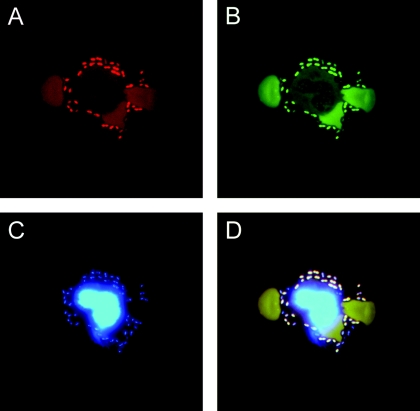
FIG. 2.
FISH detection of pneumococci in a CSF sample from a patient. The sample was stained by an S. pneumoniae-specific Cy3-labeled probe (Spn) (A) in combination with the FITC-labeled eubacterial probe (EUB 338) (B). As a further control, the DNA stain DAPI was implemented, which stained bacteria as well as the nucleus of a granulocyte (C). The overlay (D) demonstrates that the bacteria fluoresce in all channels, resulting in a white color, while the weak autofluorescence of the erythrocytes in the FITC and Cy3 channel results in a brown color.

