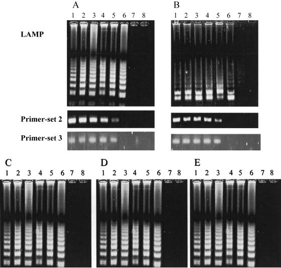
FIG. 3.
Comparative sensitivities of LAMP and PCR for the detection of HSV-1 (A) and HSV-2 (B). Amplification by LAMP shows a ladder-like pattern, whereas that by primer set 2 and primer set 3 shows single bands for the amplification products obtained by using the primer set shown in Table 2 and that from a commercial PCR kit, respectively. (C to E) Comparative sensitivities to single nucleotide polymorphisms of three HSV-1 strains. The electrophoretic profiles of the G1-4 strain (C), the conj10 strain (D), and the KH15 strain (E) are shown. Lanes: 1, 106 copies/tube; 2, 105 copies/tube; 3, 104 copies/tube; 4, 103 copies/tube; 5, 102 copies/tube; 6, 101 copies/tube; 7, 100 copies/tube; 8, negative control without target DNA.

