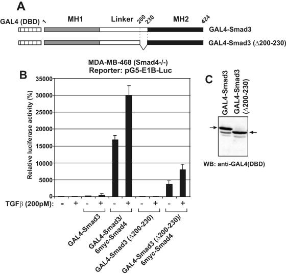
Figure 3.
Deletion of the middle region of Smad3 reduces transactivation in GAL4 assays: (A) Schematic representation of wild-type Smad3 and the Smad3 Δ200–230 mutant, fused with the DBD of the yeast transactivator GAL4 at their N-terminus. Vertical dashed lines and numbers show the coordinates of the internal deletion. (B) MDA-MB-468 cells were transfected with GAL4-Smad3 (wt) or GAL4-Smad3 (Δ200–230) vectors and the pG5-E1B-Luc reporter plasmid in the absence or in the presence of an expression vector for 6myc-Smad4 in the presence or in the absence of 200 pM of TGFβ as indicated at the bottom of the graph. The relative, normalized, luciferase activity (±SEM) is presented in the form of a bar graph. (C) Immunoblotting analysis of transfected wild-type and Δ200–230 GAL4-Smad3 proteins using an anti-GAL4 (DBD) antibody. The arrows show the position of the GAL4-Smad3 proteins.

