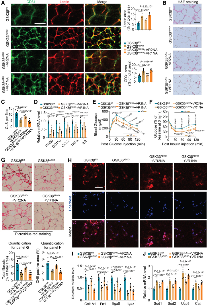
Figure 6.
VEGFR2 mediates the improvement of vascularity, metabolic complications, and the tissue microenvironment in obese adipose tissue resulting from adipocyte-specific ablation of GSK3β (glycogen synthase kinase-3 beta). Male GSK3βfl/fl and GSK3βADKO mice were fed a high-fat diet to induce obesity. The induced obese GSK3βADKO mice were then treated with either IgG or VEGFR1- (GSK3βADKO+VR1NA) or VEGFR2-neutralizing antibodies (GSK3βADKO+VR2NA) for use in the experiments. A, Representative image of epididymal white adipose tissue (eWAT) whole-mount staining of lectin and CD31 from the indicated obese mice injected with lectin and quantification of the lectin-perfused area and CD31-positive area (n=6). B and C, Hematoxylin and eosin staining of eWAT (B) and crown-like structure [CLS] quantification (C) in the indicated obese mice (n=6) using ImageJ software. D, The expression of inflammation marker genes in eWAT was determined using reverse transcription polymerase chain reaction (RT-PCR; n=6). E and F, Glucose tolerance test (E) and insulin tolerance test (F) in the indicated mice (n=6). G, Representative picrosirius red staining and its quantification for fibrosis analysis of eWAT in the indicated mice (n=6). H, Reactive oxygen species staining (dihydroethidine [DHE] dye) of eWAT sections from the indicated mice and quantification using ImageJ software (n=6). I and J, Expression analysis using RT-PCR for fibrosis-associated genes (I) and antioxidative stress-associated genes (J; n=6). Scale bar=100 µm. A, C, D, and G through I, One-way ANOVA and Tukey post hoc test; E and F, repeated ANOVA and Bonferroni post hoc test. VR1NA indicates VEGFR1-neutralizing antibody; and VR2NA, VEGFR2-neutralizing antibody.

