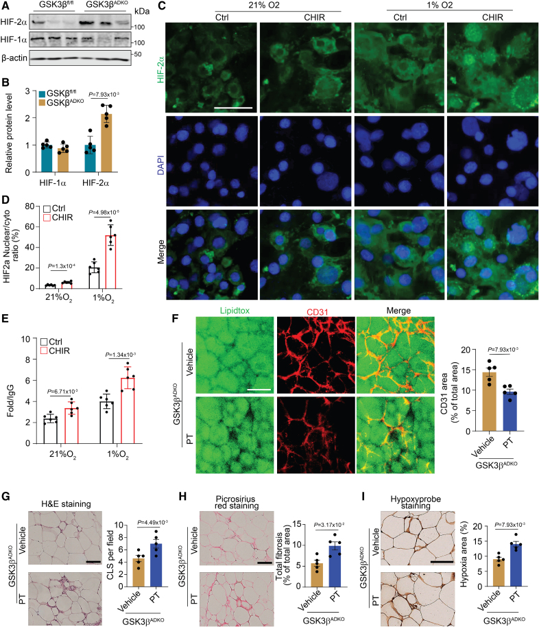
Figure 7.
The effects of adipocyte-specific GSK3β (glycogen synthase kinase-3 beta) ablation on expanded tissue vascularity and improved microenvironment of obese adipose tissue are mediated by HIF-2α (hypoxia-inducible factor 2-alpha). Male GSK3βfl/fl and GSK3βADKO mice were fed a high-fat diet to induce obesity. The induced obese GSK3βADKO mice were then treated with or without the HIF-2α inhibitor PT2385 (GSK3βADKO+PT [30 mg/kg]) for 2 weeks for use in the experiments of A and B, and F through I. A and B, Expression analysis of HIF-1α and HIF-2α at the protein level using Western blotting (A) in epididymal white adipose tissue (eWAT) of indicated obese mice and quantification using ImageJ software (B; n=5). C, Nuclear accumulation of HIF-2α in mature 3T3-L1 adipocytes, with or without GSK3 inhibitor CHIR99021 (CHIR, 10 µmol/L) treatment, under hypoxic (1%) and normoxia (21%) conditions for 4 hours. The nuclei were visualized using 4′,6-diamidino-2-phenylindole (DAPI) staining. D, Analysis of HIF-2α nuclear/cytoplasmic ratio from the experiments presented in C (n=6). E, Chromatin immunoprecipitation (ChIP) analysis of HIF-2α at the VEGF-hypoxia response element after 4 hours of 1% O2 treatment with GSK3 inhibition (n=5). F, Whole-mount CD31 staining and CD31-positive area quantification of eWAT sections from the indicated obese mice (n=5). G, Hematoxylin and eosin staining of eWAT and CLS quantification in the indicated obese mice (n=5) using ImageJ software. H, Representative picrosirius red staining and quantification for the fibrosis analysis of eWAT from the indicated obese mice (n=5). I, Representative images of hypoxyprobe staining of eWAT and quantification of staining in the indicated mice (n=5). Scale bar=100 µm. B through E, Two-tailed unpaired t test; F through I, nonparametric test.

