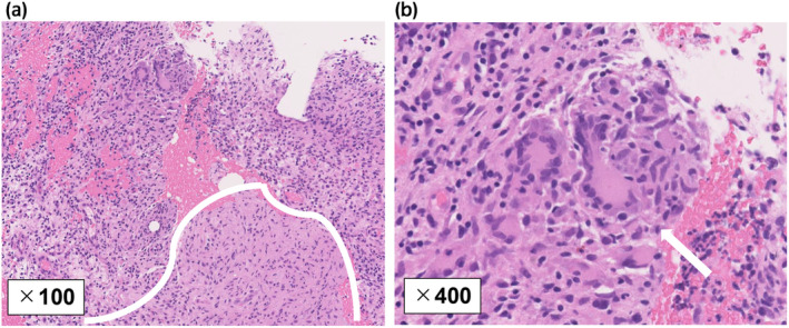
Fig. 3.

Histopathological findings for the needle biopsy. (a, b) Hematoxylin and eosin staining showed an epithelioid granuloma area (surrounded by a white line) containing Langhans giant cells (white arrow) and infiltration of inflammatory cells (a, ×100 magnification; b, ×400 magnification).
