Fig. 2.

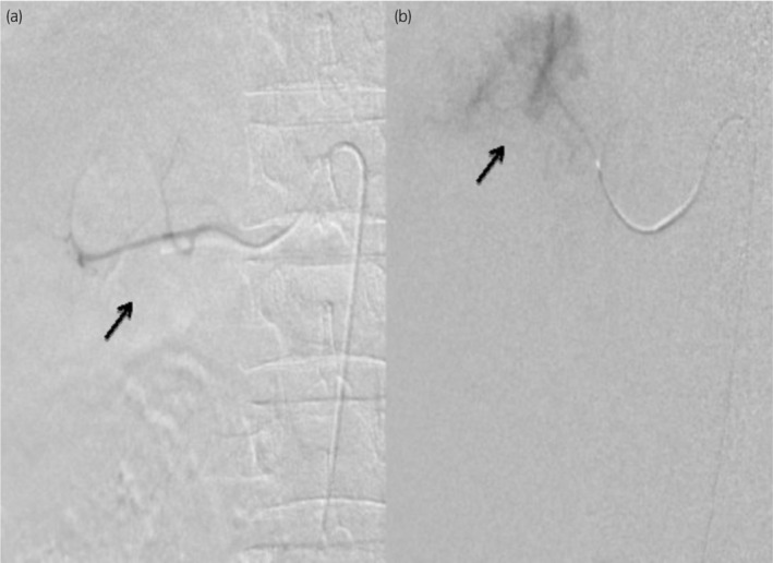
Digital subtraction angiography images showcasing the right renal artery at both midpole (a) and upper‐pole (b) regions, performed using a 5F SIMS catheter.

Digital subtraction angiography images showcasing the right renal artery at both midpole (a) and upper‐pole (b) regions, performed using a 5F SIMS catheter.