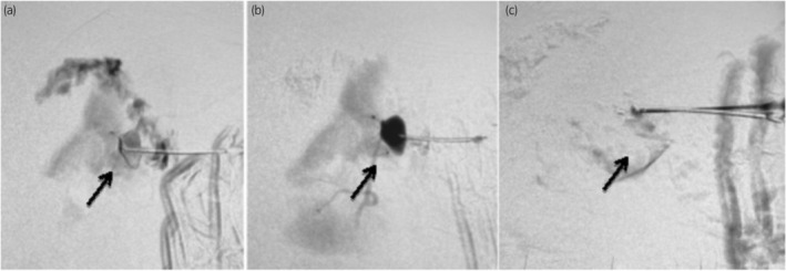
Fig. 3.

Simultaneous percutaneous puncture of the pseudoaneurysm under sonographic guidance viewed concurrently in digital subtraction angiography (DSA) (a), followed by DSA depiction of the contrast‐filled pseudoaneurysm (b), and, finally, DSA showing the absence of opacification in the pseudoaneurysm following N‐butyl cyanoacrylate glue injection (c).
