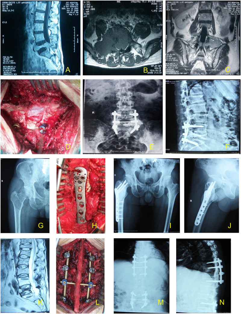
Figure 6.
The case of a patient undergoing triple sugery. The first surgery is represented by (A–F). (A–C) Prior to surgery, the lesion was located using high-resolution magnetic resonance imaging. (D) Surgical field during the first surgery. (E,F) Following surgery, the lesion was located by high-resolution magnetic resonance imaging. The second surgery is represented by (G–J). (G) MRI of patient prior to the second surgery. (H) Surgical field during the second surgery. (I,J) Following the second surgery, the lesion was located by high-resolution magnetic resonance imaging. The third surgery is represented by (K–N). (K) MRI of patient prior to the third surgery. (L) Surgical field during the third surgery. (M,N) Following the third surgery, the lesion was located by high-resolution magnetic resonance imaging. MRI, magnetic resonance imaging.

