
Figure 2.
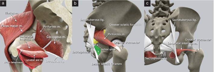
Pelvic sidewall muscles. (a) Schematic diagram of right hemipelvis, medial view. The arcus tendineus is a thickening of the fascia of the obturator internus muscle that serves as the origin of the iliococcygeus muscle. (b) Schematic diagram demonstrating insertion of piriformis muscle on apex of the greater trochanter of the femur and location of greater (yellow) and lesser (green) sciatic foramina (lateral view of right os coxae). The piriformis muscle divides the greater sciatic foramen into supra‐ and infrapiriform foramina. (c) Schematic diagram demonstrating insertion of obturator muscle onto the greater trochanter of the femur in dorsal view. lig., ligament; m., muscle.