Abstract
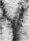
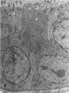
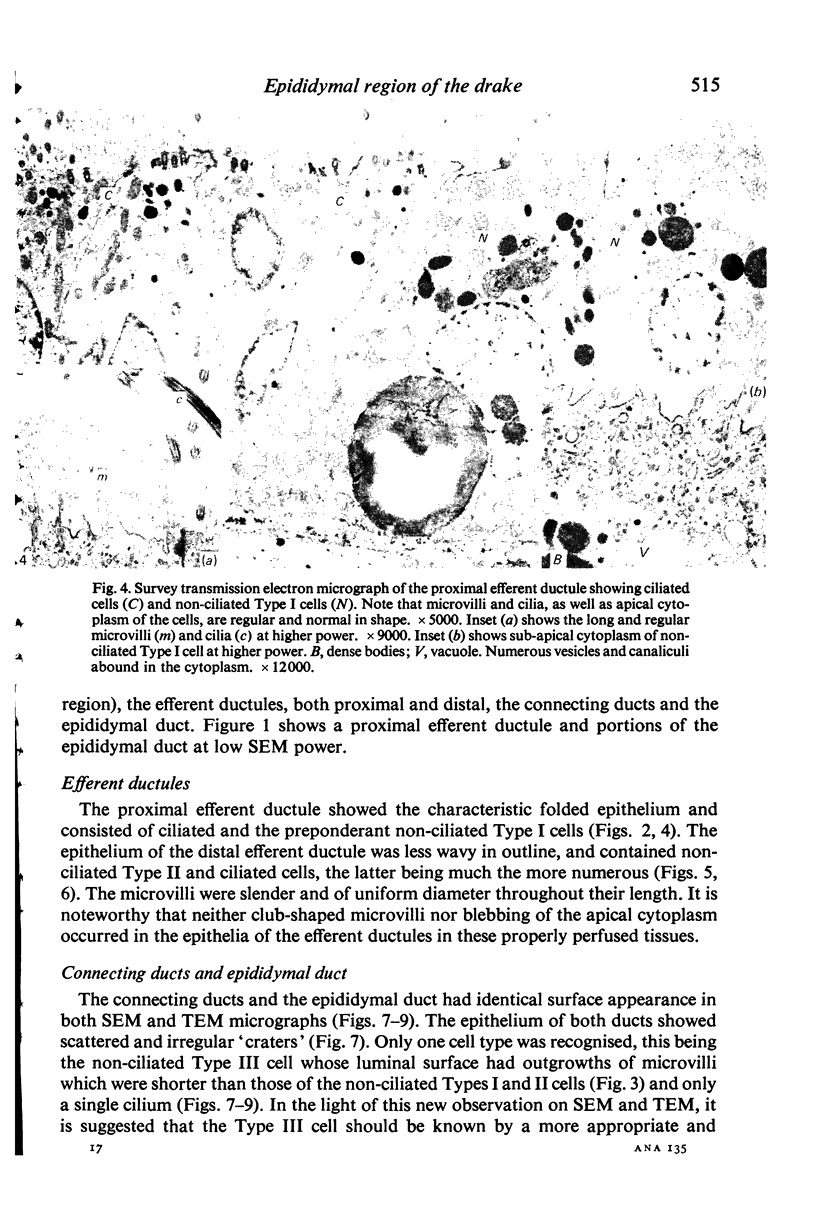
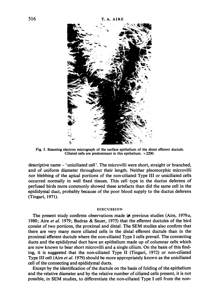
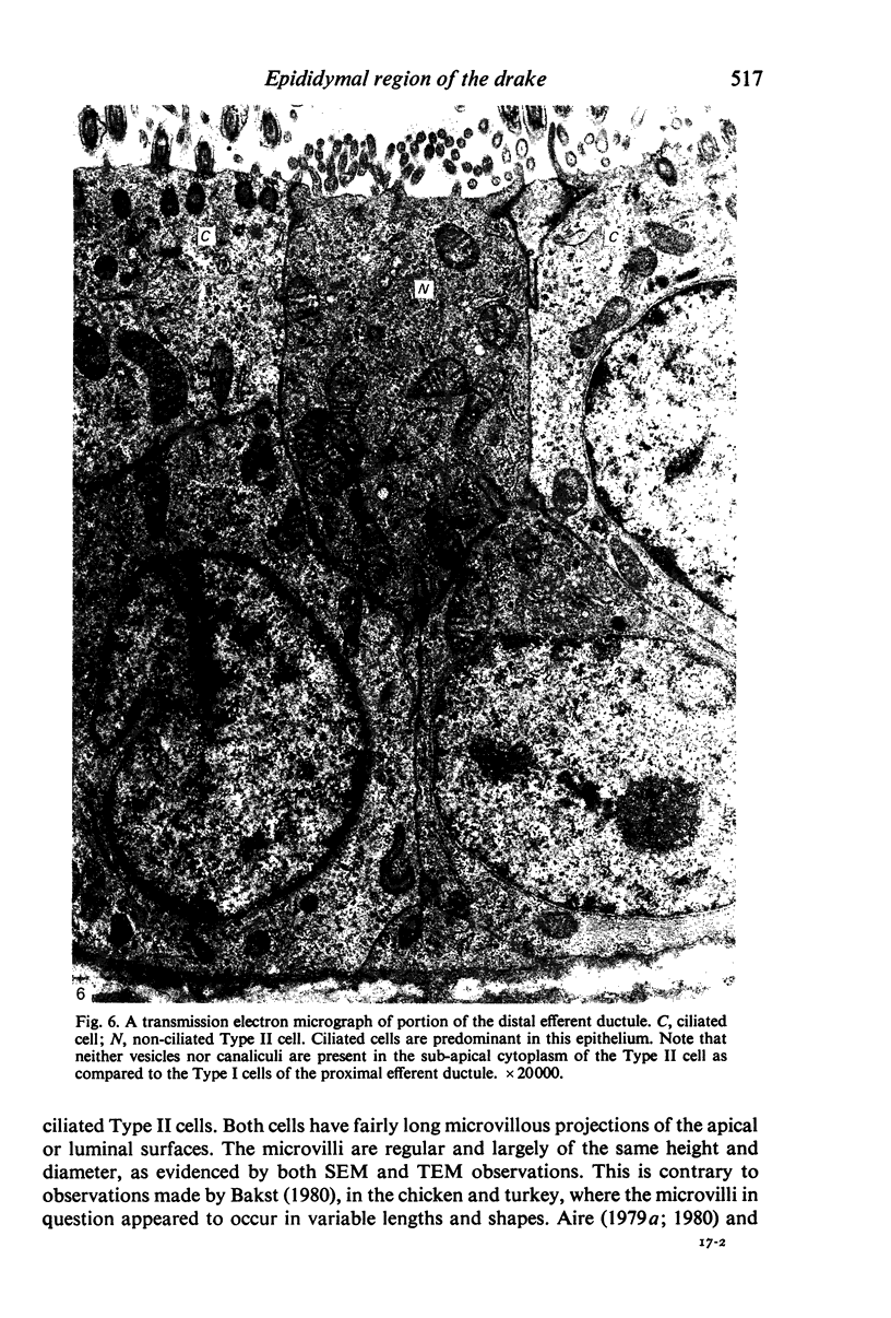
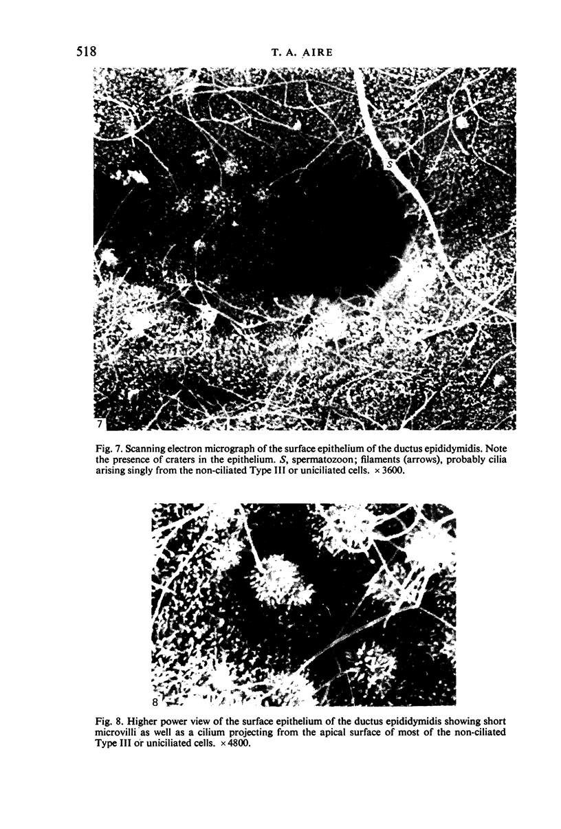
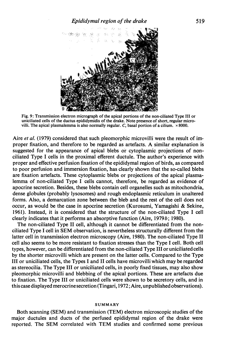
Both scanning (SEM) and transmission (TEM) electron microscopic studies of the major ductules and ducts of the perfused epididymal region of the drake were reported. The SEM correlated with TEM studies and confirmed some previous observations that the non-ciliated Types I and II cells in the proximal and distal efferent ductules, respectively, possessed apical microvilli as distinct from the cilia of the ciliated cells. The relative number of each cell type in each duct was also revealed. All microvilli and cilia were regular in shape. The connecting and epididymal ducts showed 'craters' scattered over their entire epithelial surfaces. Also, a single cilium projected from most of the cells of the epithelial lining into the lumen of these ducts. The name, 'uniciliated cell' has been suggested to describe this cell which has, until now, been referred to as the non-ciliated Type III cell (Aire, 1980; Aire et al. 1979). Neither bulbous microvilli nor blebbing of the apical plasmalemma of the cells occurred in properly fixed tissues.
Full text
PDF
Images in this article
Selected References
These references are in PubMed. This may not be the complete list of references from this article.
- Aire T. A., Ayeni J. S., Olowo-Okorun M. O. The structure of the excurrent ducts of the testis of the guinea-fowl (Numida meleagris). J Anat. 1979 Oct;129(Pt 3):633–643. [PMC free article] [PubMed] [Google Scholar]
- Aire T. A. Micro-stereological study of the avian epididymal region. J Anat. 1979 Dec;129(Pt 4):703–706. [PMC free article] [PubMed] [Google Scholar]
- Aire T. A. The ductuli efferentes of the epididymal region of birds. J Anat. 1980 Jun;130(Pt 4):707–723. [PMC free article] [PubMed] [Google Scholar]
- Aire T. A. The epididymal region of the Japanese quail (Coturnix coturnix japonica). Acta Anat (Basel) 1979;103(3):305–312. [PubMed] [Google Scholar]
- Bakst M. R. Luminal topography of the male chicken and turkey excurrent duct system. Scan Electron Microsc. 1980;(3):419–425. [PubMed] [Google Scholar]
- Budras K. D., Sauer T. Morphology of the epididymis of the cock (Gallus domesticus) and its effect upon the steroid sex hormone synthesis. I. Ontogenesis, morphology and distribution of the epididymis. Anat Embryol (Berl) 1975 Dec 23;148(2):175–196. doi: 10.1007/BF00315268. [DOI] [PubMed] [Google Scholar]
- Hess R. A., Thurston R. J., Biellier H. V. Morphology of the epididymal region and ductus deferens of the turkey (Meleagris gallopavo). J Anat. 1976 Nov;122(Pt 2):241–252. [PMC free article] [PubMed] [Google Scholar]
- Hess R. A., Thurston R. J. Ultrastructure of epithelial cells in the epididymal region of the turkey (Meleagris gallopavo). J Anat. 1977 Dec;124(Pt 3):765–778. [PMC free article] [PubMed] [Google Scholar]
- KUROSUMI K., YAMAGISHI M., SEKINE M. Mitochondrial deformation and apocrine sectetory mechanism in the rabbit submandibular organ as revealed by electron microscopy. Z Zellforsch Mikrosk Anat. 1961;55:297–312. doi: 10.1007/BF00384325. [DOI] [PubMed] [Google Scholar]
- Tingari M. D. On the structure of the epididymal region and ductus deferens of the domestic fowl (Gallus domesticus). J Anat. 1971 Sep;109(Pt 3):423–435. [PMC free article] [PubMed] [Google Scholar]


