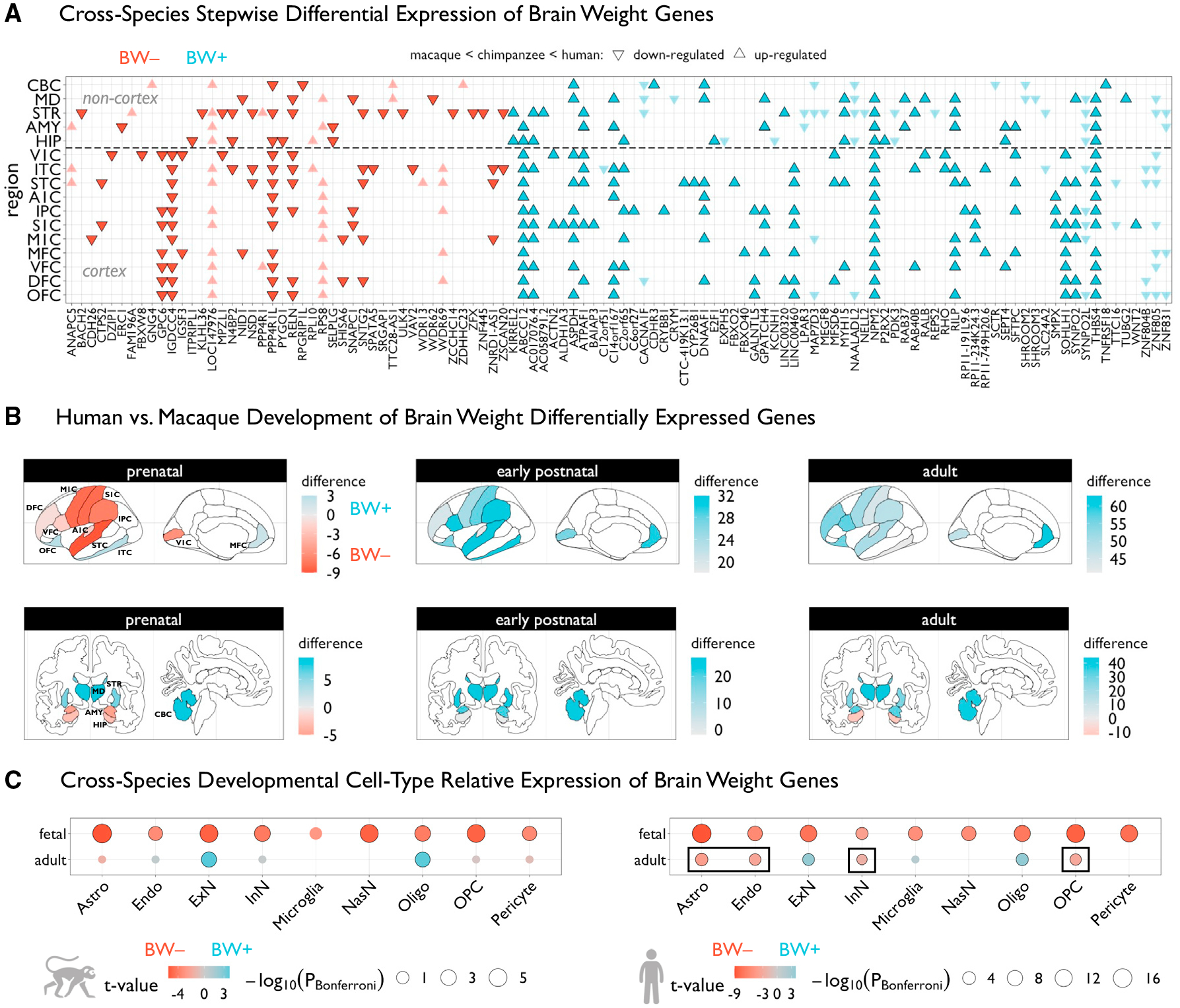
Figure 2. Genes associated with BW show differential effects across species.

(A) Grid plot showing BW-associated genes that are also significantly differentially expressed in humans relative to chimpanzees and macaques (pBonferroni < 0.05), across 16 brain regions (11 neocortical areas). These are BW-associated genes that were previously defined42 as having higher or lower expression compared with the two species collectively, and reanalyzed to assess stepwise interspecies effects that reflect the absolute differences in brain size between species (i.e., up-regulated implies gene expression humans > chimpanzees > macaques, while downregulated implies gene expression humans < chimpanzees < macaques). Triangles represent directions of effects and colors denote the respective BW gene set. Genes that are highlighted show congruent directional effects in respective BW sets (i.e., BW+ and upregulated in humans, and vice versa).
(B) Brain plots of differences in counts of number of BW+ or BW− genes that were defined to be significantly differentially expressed in humans relative to macaques each of three developmental epochs—a positive regional difference (blue) indicates that BW+ genes tend to be upregulated in humans in that region in a given developmental epoch, while a negative difference (red) indicates that BW− genes tend to be upregulated. The same 16 regions from (A) are shown anatomically, based on manual assignment using a common human atlas.44
(C) Differential expression of BW+ versus BW− genes across individual cell types, using cell-specific RNA sequencing data in fetal and adult samples from macaques (left) and humans (right). BW− relative expression (red) indicates that BW− genes are more highly expressed in that cell type compared with BW+, whereas BW+ relative expression (blue) indicates the opposite effect. Black outlines denote significant effects (pBonferroni < 0.05). Circles are scaled according to Bonferroni-corrected p values. Black rectangles denote human-specific effects relative to macaques. V1C, primary visual cortex; M1C, primary motor cortex; S1C, primary somatosensory cortex; A1C, primary auditory cortex; ITC, inferior temporal cortex; IPC, inferior parietal cortex; STC, superior temporal cortex; OFC, orbitofrontal cortex; VFC, ventrolateral frontal cortex; DFC, dorsolateral frontal cortex; MFC, medial frontal cortex; STR, striatum; MD, medial dorsal thalamus; AMY, amygdala; HIP, hippocampus; CBC, cerebellar cortex. Astro, astrocytes; Endo, endothelial cells; ExN, excitatory neurons; InN, inhibitory neurons; NasN, nascent neurons; Oligo, oligodendrocytes; OPC, oligodendrocyte precursor cells.
