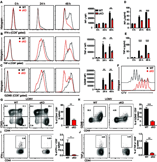
Fig. 1.

PP2A deficiency inhibits CD8+ T cell effector function. (A to C) WT or PP2A-deficient CD8+ T cells were stimulated with anti-CD3 (0.1 ng/μl) plus anti-CD28 (3 ng/μl) for the specified durations (0, 24, and 48 h). IFN-γ, TNF-α, and GZMB expression were measured by flow cytometry (intracellular cytokine staining). Representative images of IFN-γ, TNF-α, and GZMB intracellular staining and mean fluorescence intensity of the stained cells are presented. (D and E) Supernatant from (A) to (C) was harvested and assayed for IFN-γ and TNF-α by ELISA. (F) CTV-labeled naïve CD8+ lymphocytes were isolated from WT and Ppp2cafl/fl/dLckcre mice and stimulated for 3 d. Flow cytometry was used to assess the dilution of CTV on the CD8+ T cells. (G) Splenocytes from the LCMV-infected mice (5 d after infection) were isolated and gated on CD8+ T cells. Representative plots depicting the number of CD44+ CD8+ T cells (left) and their percentage (right) among the splenocytes of mice of different types (n = 3). (H to J) Isolated splenocytes from (G) were gated on CD8+ T cells. Representative plots (left) and the frequency (right) of GZMB+ CD44+ (H), IFN-γ+ CD44+ (I), and TNFα+ CD44+ (J) among CD8+ T cells are presented (n = 3). Mean ± SEM. *P < 0.05, **P < 0.01, ***P < 0.001, 2-tailed unpaired Student’s t test.
