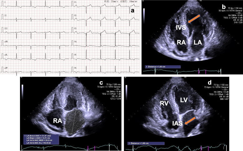
Fig. 1.
Electrocardiogram and echocardiogram features. a ECG documented that the patient had sinus bradycardia, first degree atrioventricular block, and myocardial ischemia. b TTE demonstrated left and right ventricular myocardial hypertrophy, the myocardium echoed like "ground glass" (yellow arrow). c From apical four-chamber view, TTE detected the dilated left and right atrium. d TTE showed the thickened interatrial septum (yellow arrow) in apical four-chamber view. ECG: electrocardiogram; TTE: transthoracic echocardiography; LA: left atrium; RA: right atrium; IVS: interventricular septum; IAS: interatrial septum

