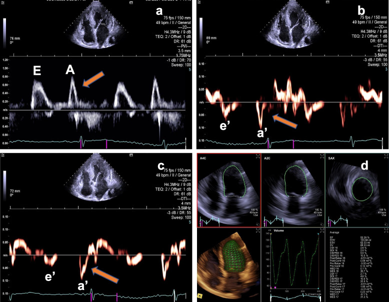
Fig. 2.
TTE and RT4D-TTE revealed the left ventricular ejection fraction and diastolic function. a Pulse wave Doppler showed that peak A velocity was higher than peak E of the mitral flow (yellow arrow). b Tissue Doppler Imaging (TDI) detected peak a’ velocity was higher than peak e’ of the mitral annular (yellow arrow). c Representative images of LV strain values, STE of VVI technique detected the patient with a GLS of −9.00%. d Automatic volumetric left ventricular analysis (LVA) showed that LV ejection fraction was 52.28%. RT4D-TTE: real-time 4-dimensional transthoracic echocardiography; TDI: Tissue Doppler Imaging

