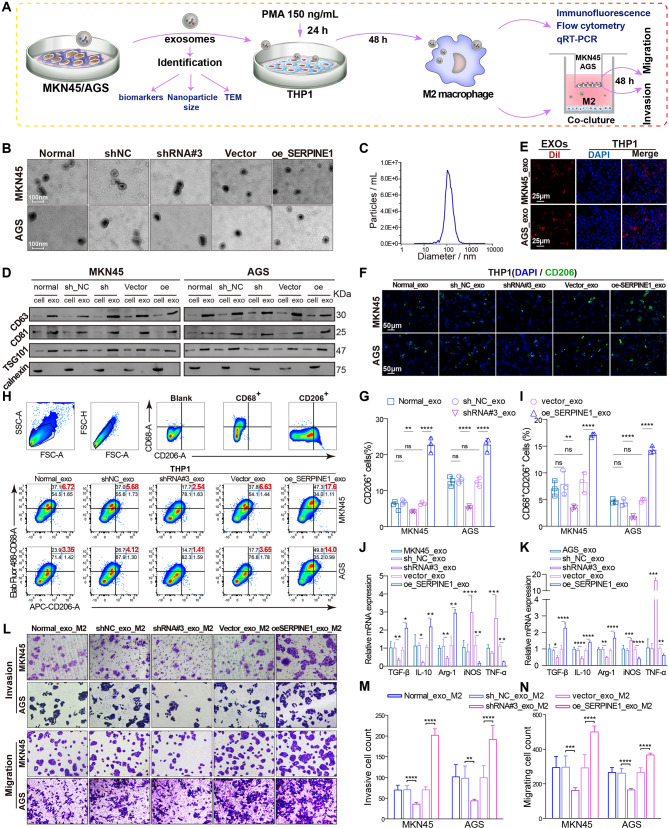
Fig. 5.
SERPINE1-mediated gastric cancer-derived exosomes facilitate the polarization of THP1 cells into M2 macrophages. (A) Schematic representation of the extraction and identification of exosomes and the induction of macrophage polarization. Transmission electron microscopy (B), nanoparticle tracking analysis (C), and western blotting (D) were used to identify the morphology, particle size, and markers of exosomes. (E) Confocal laser scanning microscopy detected Dil-labeled exosomes (red) internalized by DAPI-labeled macrophages (blue). (F–G) Immunofluorescence analysis of the proportion of CD206+ cells in THP1 cells treated with exosomes. (H–I) Flow cytometry analysis of the proportion of CD68+CD206+ cells in THP1 cells treated with exosomes. (J–K) qRT-PCR analysis of M1 markers (iNOS and TNF-α) and M2 markers (TGF-β, IL-10, and Arg-1) in THP1 cells treated with exosomes. (L–N) Transwell migration and invasion assays of GC cells (upper chamber) co-cultured with macrophages (lower chamber) ingesting exosomes

