Figure 5.

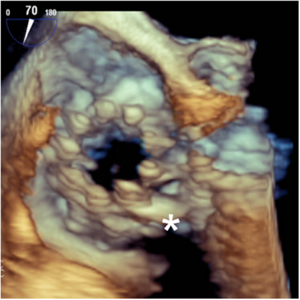
3D‐ Transesophageal echocardiographic image of the implanted valve. The XTW Triclip is being pushed toward the valvular anulus (asterisk). [Color figure can be viewed at wileyonlinelibrary.com]

3D‐ Transesophageal echocardiographic image of the implanted valve. The XTW Triclip is being pushed toward the valvular anulus (asterisk). [Color figure can be viewed at wileyonlinelibrary.com]