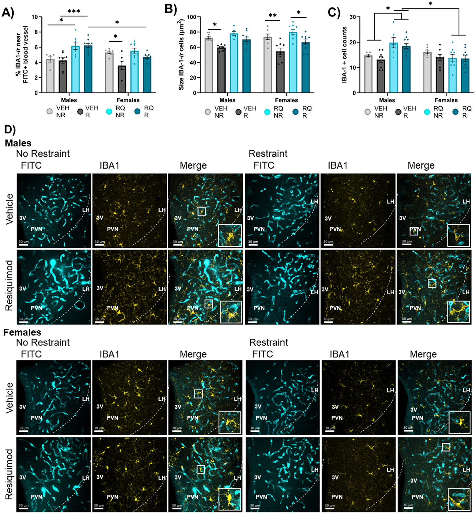
Fig. 4. Microglia localization, size, and number differed among offspring of prenatally injected mothers.

(A) Proximity of microglia to blood vessels (within 1.18 μm), (B) average size of IBA-1-labeled microglia cells, and (C) the number of IBA-1-labeled cells in the PVN. (D) Representative images. VEH = vehicle, RQ = Resiquimod, NR = no restraint, R = restraint, PVN = Paraventricular Nucleus of the Hypothalamus, 3 V = 3rd ventricle, FITC = fluorescein isothiocyanate, IBA1 = ionizing binding calcium adapter molecule-1. *p < 0.05, **p < 0.01, ***p < 0.001, ****p<0.0001. Error bars represented as +/− SEM. n = 8–10 animals / group.
