
Figure 1. Paraxial mesoderm segmentation and modules of somitogenesis.
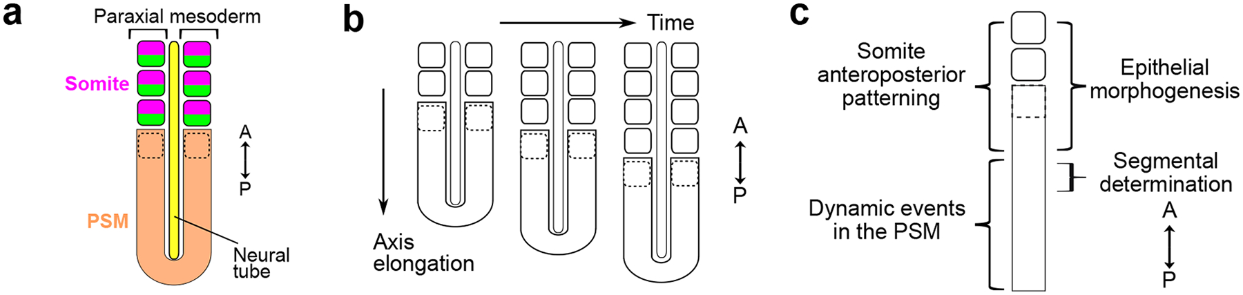
a. The paraxial mesoderm locates on either side of the neural tube. It forms epithelial blocks called somites from the unsegmented region called presomitic mesoderm (PSM). Each somite is subdivided into an anterior (magenta) and posterior (green) compartment associated with different transcriptomes and developmental trajectories. The dotted outline represents the forming somite. A, anterior; P, posterior.
b. Somites are formed sequentially in an anterior to posterior direction, in coordination with the elongation of the body axis.
c. Simplified representation of the paraxial mesoderm and general locations of different somitogenesis modules. In the anterior PSM, both somite anteroposterior patterning and epithelial morphogenesis are initiated with the expression of MESP transcription factors, just anteriorly to segmental determination.