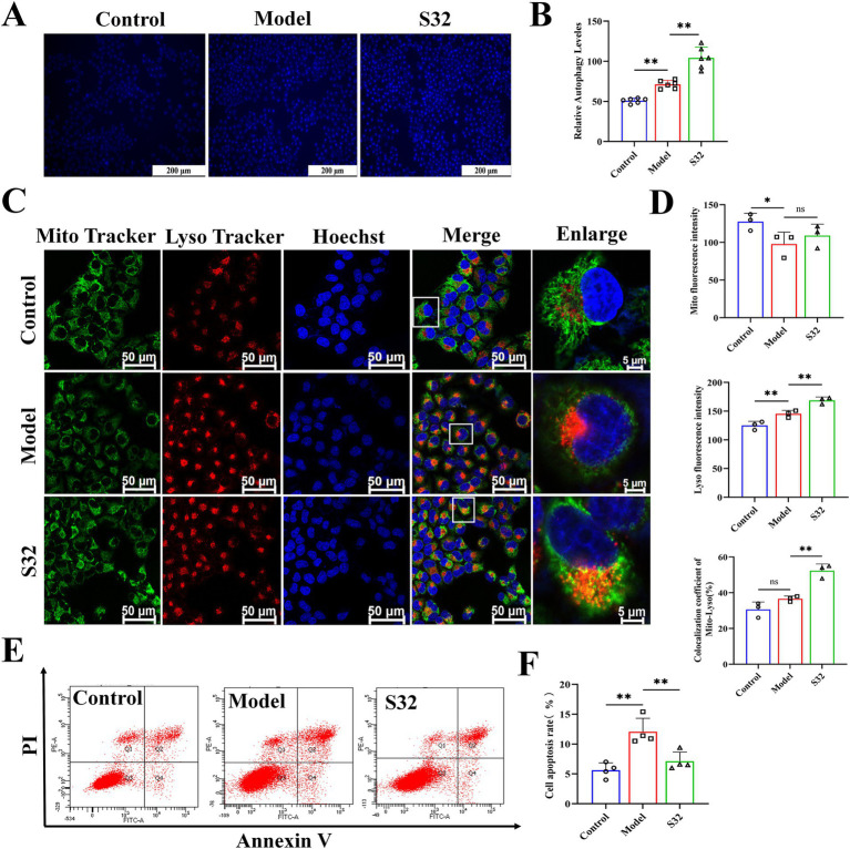
Figure 4.
Sesamin activates mitophagy and reduces apoptosis in L02 cells treated by 9-trans-C18:1. (A,B) Semiquantitative measurement of intracellular MDC levels and fluorescence intensity; (C) Fluorescence intensity photography of Mito-Lyso co-localization; (D) Mitochondrial fluorescence intensity, semiquantitative lysosome fluorescence intensity, and Mito-Lyso red green fluorescence co-localization coefficient (n ≥ 50 cells per group); (E,F) Apoptosis rate and semiquantitative analyses; (A) Scale bar = 200 μm, (C) scale bar = 50 μm. Control: RPMI 1640 complete culture medium; Model: 500 μM 9-trans-C18:1 + RPMI 1640; S32: 500 μM 9-trans-C18:1 + 32 μM sesamin. All data are represented as means ± SD (n = 3–6), *p < 0.05; **p < 0.01.

