Abstract
Ehrlichia ruminantium, an obligate intracellular bacterium transmitted by ticks of the genus Amblyomma, causes heartwater disease in ruminants. The gene coding for the major antigenic protein MAP1 is part of a multigene family consisting of a cluster containing 16 paralogs. In the search for differentially regulated genes between E. ruminantium grown in endothelial and tick cell lines that could be used in vaccine development and to determine if differences in the map1 gene cluster exist between different isolates of E. ruminantium, we analyzed the map1 gene cluster of the Senegal and Gardel isolates of E. ruminantium. Both isolates contained the same number of genes, and the same organization as found in the genome sequence of the Welgevonden isolate (H. Van Heerden, N. E. Collins, K. A. Brayton, C. Rademeyer, and B. A. Allsopp, Gene 330:159-168, 2004). However, comparison of two subpopulations of the Gardel isolate maintained in different laboratories demonstrated that recombination between map1-3 and map1-2 had occurred in one subpopulation with deletion of one entire gene. Reverse transcription-PCR on E. ruminantium derived mRNA from infected cells using gene-specific primers revealed that all 16 map1 paralogs were transcribed in endothelial cells. In one vector (Amblyomma variegatum) and several nonvector tick cell lines infected with E. ruminantium, transcripts were found for between 4 and 11 paralogs. In all these cases the transcript for the map1-1 gene was detected and was predominant. Our results indicate that the map1 gene cluster is relatively conserved but can be subject to recombination, and differences in the transcription of map1 multigenes in host and vector cell environments exist.
Ehrlichia ruminantium (formerly Cowdria ruminantium [12]) is the causative agent of heartwater, a rickettsial disease transmitted by ticks of the genus Amblyomma which causes major economic losses in wild and domestic ruminants. The disease is endemic in sub-Saharan Africa and also is present on some Caribbean islands (33), where it poses a risk of spreading to the American mainland. Feeding ticks transmit E. ruminantium to vertebrate hosts in their saliva and/or by gut regurgitation (8, 19). Phylogenetic studies have revealed a close relationship between E. ruminantium, Ehrlichia canis, and Ehrlichia chaffeensis (30, 39).
In infections with these ehrlichial agents, the serological response is mainly directed against outer-membrane proteins of approximately 30 kDa. The genes coding for these proteins have been designated the major antigenic protein 1 (map1) in E. ruminantium (32, 40), the outer membrane protein p28 (omp-1) in E. chaffeensis, and the p30 outer membrane protein (p30) in E. canis (27, 28, 30, 35, 36, 42, 43). The OMP-1 and P30 protein families are each encoded by a multigene family consisting of 22 genes arranged in a cluster between a hypothetical transcriptional regulator (upstream) and the secA gene (downstream). The 5′ end of the cluster contains paralogs with short intergenic spaces, whereas the paralogs at the 3′ end are separated by larger intergenic spaces. Previously only four E. ruminantium map1 paralogs had been identified, including one located downstream from map1, designated map1+1, which was located upstream from secA (37). Very recently the whole map1 cluster was characterized for the Welgevonden isolate of E. ruminantium and shown to contain 16 genes located downstream from a hypothetical transcriptional regulator and the secA gene (38).
In vitro systems for the propagation and study of E. ruminantium using mammalian endothelial cells have been available for nearly 20 years, but systems for the propagation of E. ruminantium in tick cell lines have become available only recently (4-6). Using these in vitro systems, it was shown that there are clear differences in morphology between bacteria grown in endothelial cell and tick cell cultures (6) and also in their immunogenicity and pathogenicity for sheep (7). Together these data strongly suggest that differences may occur in gene transcription and protein expression by E. ruminantium grown in endothelial and tick cell cultures.
Various studies investigating the transcription of ehrlichial multigene families have been conducted using both in vitro cultures and in vivo material collected from infected vertebrate hosts and ticks. All 22 paralogs in the E. canis p30 multigene family and 16 paralogs in the E. chaffeensis p28 multigene family were found to be transcribed when reverse transcription-PCR (RT-PCR) was used on infected monocyte cultures using gene-specific primers (23, 27). The p30 paralogs with short intergenic regions were cotranscribed in monocyte cultures (27). In infected vertebrates, 16 out of 22 and 11 out of 14 paralogs studied were found to be transcribed for E. chaffeensis and E. canis, respectively, whereas only 1 paralog was found to be transcribed in various tick stages (13, 35, 36). We recently investigated the transcription of map1-2, map1-1, and map1 of E. ruminantium grown in bovine endothelial cells, tick cell lines, and in Amblyomma variegatum ticks (2). In that study we found that map1 was always transcribed and map1-1 was transcribed in tick cells in vitro, ticks in vivo, and by attenuated organisms in endothelial cells, but we could not detect a transcript for map1-2 in any of the studied samples. However, van Heerden et al. (38) showed that all 16 map1 paralogs were transcribed in infected endothelial cell cultures.
To study whether all 16 E. ruminantium map1 paralogs were also transcribed in a tick environment, we studied the transcription in one vector and several nonvector tick cell lines. In order to be able to do this, the map1 multigene sequence of two isolates was determined. The study led to the identification of recombination between two map1 paralogs in one of the E. ruminantium isolates and demonstrated the occurrence of differential transcription of E. ruminantium map1 paralogs. Analysis of differential transcription of map1 multigenes in host and vector will aid in understanding the role these genes (and their products) may play in the pathogenesis and transmission of heartwater.
MATERIALS AND METHODS
Growth of E. ruminantium in bovine endothelial cells and tick cell lines.
The Gardel (34), Senegal (18), and Welgevonden (11) isolates of E. ruminantium were cultured in bovine pulmonary artery (BPC), aorta endothelial (BAE), or umbilical cord (BUE) cells as described previously (17, 26). Two subpopulations of the Gardel isolate were used; both originated from CIRAD-EMVT, Guadeloupe. One culture, designated IBET Gardel, had been transferred to the Instituto de Biologia Experimental e Tecnologica, Oeiras, Portugal, in 2001, and thence to Utrecht University in 2002. The other culture, designated CTVM Gardel, was transferred to the Centre for Tropical Veterinary Medicine (CTVM), University of Edinburgh, Scotland, in 1993. For RNA extraction the following cultures were used: CTVM Gardel in BPC between passages 12 and 15, IBET Gardel in BAE between passages 45 and 66, Senegal in BAE between passages 4 and 14, and Welgevonden in BAE between passages 7 and 16. The CTVM Gardel and Welgevonden isolates of E. ruminantium were maintained in vitro in tick cell lines at temperatures between 28°C and 37°C as described previously (4-6). Both isolates were grown in the following cell lines: Ixodes scapularis embryo-derived IDE8 (25), Amblyomma variegatum larva-derived AVL/CTVM13 (6), and Rhipicephalus appendiculatus nymph-derived RAN/CTVM3 (2). CTVM Gardel was also grown in BDE/CTVM16, BME/CTVM2, and IRE/CTVM18 derived from embryos of Boophilus decoloratus, Boophilus microplus, and Ixodes ricinus, respectively (4), and Welgevonden was also grown in two other R. appendiculatus lines: embryo-derived RAE/CTVM1 (3) and neonate larva-derived RAE25 (20). Bacterial growth was monitored in Giemsa-stained cytocentrifuge smears.
PCR amplification, cloning, and sequencing of the map1 multigene family.
For the Senegal isolate, the Ehrlichia canis p30 multigene family sequence (accession number AF078553) and the E. chaffeensis p28 multigene family sequence (accession numbers U72291 and AF021338) were used to do BlastN searches in the E. ruminantium genome database of the Welgevonden isolate (Sanger Centre; www.sanger.ac.uk/Projects/Microbes/). Single-hit runs were aligned where possible, and several primers were designed to amplify the E. ruminantium map1 locus. Using a Long and Accurate PCR (LA-PCR) kit (Takara Biomedical, Ohtsu, Japan), three PCR products (Fig. 1) were generated from genomic DNA extracted from E. ruminantium (Senegal) grown in bovine endothelial cells using the following PCR conditions: initial denaturation of 4 min at 94°C followed by 35 cycles of 20 s at 98°C, 10 s at 55°C, 15 min at 68°C, and a final extension of 10 min at 72°C. The PCR products were cloned into the pGEMT-easy vector (Promega Benelux, Leiden, The Netherlands) following the manufacturer's instructions and were transferred to Escherichia coli JM109 cells. Positive clones were selected by PCR amplification of part of the insert. Three overlapping clones were selected, and the sequence of the inserts was determined (Baseclear, Leiden, The Netherlands). Both primer walking and random shotgun cloning were used to determine the sequences.
FIG. 1.
Schematic representation of the E. ruminantium Senegal map1 multigene family. The genes and their orientations are represented as arrows. The bars indicate the three clones that were constructed with long-template PCR. The map1 paralog names are indicated below the solid arrows and the other gene names below the open arrows. htr, hypothetical transcriptional regulator; un-1, unknown gene.
DNA and RNA isolation.
Total RNA from endothelial and tick cell cultures was extracted as described previously (2), except that DNase treatment was done after elution from the column. DNA of the various E. ruminantium isolates was extracted as described before (40) or using the tissue protocol provided with the QIAamp extraction kit (Westburg, Leusden, The Netherlands).
cDNA synthesis and paralog-specific PCR.
Random hexamer primers were used to prepare cDNA using the SuperScript first-strand synthesis system (Invitrogen, Breda, The Netherlands). Between 5 and 10 μl of DNase-treated RNA was used to prepare 40 μl of cDNA using 100 ng of random hexamer primers according to the manufacturer's instructions. Control samples were prepared by omitting reverse transcriptase from the reaction. Two microliters of cDNA was subsequently used as template in a PCR containing 1× Taq PCR buffer (Promega, Leiden, The Netherlands), 3 mM MgCl2, 1.25 U of Taq polymerase (Promega), 400 μM of each deoxynucleoside triphosphate, and 10 pmol of each primer set (Table 1) in a 25-μl reaction. Reactions were carried out on an iCycler (Bio-Rad Laboratories b.v., Veenendaal, The Netherlands) using the following program: 2 min at 94°C followed by 40 cycles of 30 s at 94°C, 30 s at 55°C, 1 min at 72°C, and a final elongation step of 7 min at 72°C. Positive controls included genomic DNA instead of cDNA. PCR products were visualized by running the samples on ethidium-bromide-stained agarose gels.
TABLE 1.
Primers used for the RT-PCR on the map1 cluster of E. ruminantium Senegal and Gardel
| Genea | Senegal (5′-3′) | Gardel (5′-3′) |
|---|---|---|
| map1-14 (f) | TGTTGACTTTTCCAATGAGAGTGA | TGTTGACTTTTCCAATGAGAGCGA |
| (r) | TTGGTGAAAGTAAAAACCCCATAC | TTGGTGAAAGTAAAAACCCTACAC |
| map1-13 (f) | AGATCTTAAAGAGGATGGGTACAA | AGATCTTAAAGAGGATGGATACAA |
| (r) | GGTAATAACCTTCTGCAACAGCTA | CTAAAATAAACCTTACTCCTATAC |
| un-1 (f) | ATTAACAGCACTCCCAATCCATTA | ATTAGCAGCACTCCTAATCCAGTA |
| (r) | AATTTGTACAGCTGCTTGGAAAAA | GGAAAACAACACTTTTTGTGGGTG |
| map1-12 (f) | AATACAAGCCAAGCATTTCGTACT | AATACAAGCCAAGCATTTCGTATT |
| (r) | TAACATTGAATTTTGCTGATGATG | TAACGTTGAATTTTGCTGATGATG |
| map1-11 (f) | TATCAAACTTTCGAAACTTCCACA | TATCAAACTTTCGAAACTTTCACA |
| (r) | CACTGATCAGGAGATTTGTTCTTG | CACTGATCAAGAGATTTGTTCTTG |
| map1-10 (f) | ATTACCACCCAATTTAAGCCTACT | ATTACCAGCCAATTTAAGCCTACT |
| (r) | AATCCTTAACCCGACTTCGCTACC | AATCCTTAACCCGACTTCGCTACC |
| map1-9 (f) | GTAACGTACCAAATACTGAAGATG | GTAACGTACCAAATACTGAAGATG |
| (r) | TATTAAGTTTGGCTATTGCTGAA | TATTAAGTTTGGCTATTGCTGAAG |
| map1-8 (f) | TTTAACATTCTTATTAGCGTTTTC | TTTAACATTCTTATTAGCGTTCTC |
| (r) | CTGATTTTAGGAGATACGCGATAA | CTGATTTTAGGAGATACACGATAA |
| map1-7 (f) | TGGACAATATAAGCCAGGAGTTCC | TGGACAATATAAGCCAGGAGTTCC |
| (r) | ACTGCACTAGTAATTTTGGGTTCA | GTTGCACTAGTAATTTTGGGTTCA |
| map1-6 (f) | TTTTTACCTCACCAAGCACTTTC | TTTTTACCTCACCAAGTACTTTC |
| (r) | AGCTAATGTTTAACGTTGATGTTG | AGCTAATGTTTAATGTTGATGTTG |
| map1-5 (f) | CTTTTGCCTTCATTCATACATTTA | ATACAAAACTAGAAAAAGAGAATA |
| (r) | TACTTACTTTGACATTATATGCTA | TACTTACTTTGACATTATATGCTA |
| map1-4 (f) | GTTTCAGTGGCACTATAGGACAGT | GTTTTAGTGGCACTATTGGACAGT |
| (r) | TTGGGATTATTTGCAAGCATACGA | TTGGGATTATTTGCAAGCATACGA |
| map1-3 (f) | CAATTTTCGTAACTTCTCAGTCAA | CAATTTTCGTAATTTCTCAGTTAA |
| (r) | TTTAAATTTATTGCCTGCTACTTT | TTTAAATTTATTACCTACTACTTT |
| map1-2 (f) | AATAAACTCATTGCAACAGGTATA | AACAAACTCATTGCAACAGGTATA |
| (r) | CTAATGGGATGTTATAGAGATCAG | CTAATGGGGTGTTATTCAGATCAG |
| map1-1 (f) | CCAAGCATACCACATTTCAGA | CCAAGCATACCACACTTCAGA |
| (r) | TGAAGCGGAAGTGCTTTGAGG | TGAAGCGGAAGTGCTTTGAGG |
| map1 (f) | TAATATCATTAGTGTCATTTTTACC | TAATATCATTAGTGTCATTTTTACC |
| (r) | TGGACTAACAGCACTACTGGC | GTGTTGCTGATGCAAAACCTGG |
| map1+1 (f) | CTTAGTTAAACCAGGGATTG | ATAATCAATATCTAAATTAGCAGA |
| (r) | AGGTGTTACATTAAGTGCAT | GGTGTTACATTAAGTGCATTGTTG |
f, forward; r, reverse.
Southern blot with map1-2, map1-3, and recombination site-specific probes.
DNA from E. ruminantium Gardel (IBET or CTVM)-infected BUE cells was obtained by scraping cells from the bottom of culture flasks. The cell suspension was passaged 10 times through a 25-gauge needle and subsequently spun at 2,000 × g for 5 min at 4°C. The supernatant containing bacteria was then spun for 15 min at 15,000 × g at 4°C. The pellet was resuspended in phosphate-buffered saline (PBS) and treated with DNase (Promega Benelux, Leiden, The Netherlands) for 30 min at 37°C to digest the host cell DNA. Bacteria were washed three times with PBS, and DNA was extracted using the QIAamp kit (Westburg, Leusden, The Netherlands). DNA from uninfected cells was directly extracted with the same kit. Genomic DNA was digested with HindIII and separated in a 0.7% agarose gel. DNA was transferred to a Nylon membrane (Zeta-probe; Bio-Rad, Veenendaal, The Netherlands), and blots were prehybridized at 50°C in prehybridization solution (6×SSPE [1× SSPE is 0.18 M NaCl, 10 mM NaH2PO4, and 1 mM EDTA {pH 7.7}], 0.5% sodium dodecyl sulfate [SDS], 100 μg/ml denatured salmon sperm DNA, and 5×Denhardt's solution [1× Denhardt's solution is 0.02% Ficoll, 0.02% polyvinylpyrrolidone, and 0.02% bovine serum albumin]) for 2 h and hybridized at 50°C in prehybridization solution containing biotin-labeled probe (Isogene, Maarssen, The Netherlands). Blots were then washed twice for 10 min in 2× SSPE, 0.1% SDS at 50°C. Subsequently the membrane was incubated in 20 ml of 1:4,000-diluted peroxidase-labeled streptavidin (Boehringer, Mannheim, Germany) in 2× SSPE-0.1% SDS for 1 h at 50°C. The membrane was washed twice in 2× SSPE-0.1% SDS for 10 min at 50°C with shaking and once in 2× SSPE for 10 min at room temperature. The membrane was thereafter incubated for 1 min in 5 ml of ECL detection fluid and exposed to an ECL hyperfilm (Amersham Pharmacia Biotech, Roosendaal, The Netherlands). The membrane was stripped by two washes in 1% SDS-0.1× SSPE for 15 min at 95°C and one at 20°C. For reuse the membrane was placed between sheets of Whatman paper and sealed in a plastic bag and stored at 4°C. The 5′ biotin-labeled probes were GAAATCCAAATCCTGGACCT for map1-3, AACAAACTCATTGCAACAGGTATA for map1-2, and ACATCTGCAATAGC(G/T)ACACTT for the recombination site.
Nucleotide sequence accession numbers.
The obtained sequence for the E. ruminantium Senegal isolate map1 multigene family was deposited as GenBank accession number AF319940. For the IBET Gardel isolate the sequence of the map1 cluster (accession number AY652746) was taken from the whole-genome sequence that was obtained by random shotgun cloning (IGH-CIRAD Ehrlichia ruminantium genome project). The map1 cluster sequence of the Welgevonden isolate was obtained from the whole-genome sequence of this isolate (accession number NC_005295).
RESULTS
Sequence analysis and cluster comparison.
Sequence analysis of the map1 cluster of both the Senegal (Fig. 1) and IBET Gardel isolates revealed the same number of map1 paralogs in these isolates as described for the Welgevonden isolate (38). Furthermore, the position of the cluster in the genome (between a hypothetical transcriptional regulator gene and the secA gene) and the arrangement of the cluster were conserved among the three different isolates. All genes within the cluster contained both start and stop codons, and only the genes un-1 and map1-12 did not contain an intergenic region and instead had a 4-nucleotide overlap (Table 2). Fifteen of the paralogs were orientated in a head-to-tail direction, with small intergenic regions (8 to 39 nucleotides) near the 3′ end region (paralogs map1-12 to map1-2) and large intergenic regions (372 to 1,621 nucleotides) in the 5′ region (paralogs map1-2 to map1+1) of the cluster. Comparison of individual paralogs using ClustalV analysis on the amino acid sequence showed highly conserved paralogs (>95% similarity; e.g., map1-9, map1-8, and map1-1) and less conserved (variable) paralogs (<86% similarity; e.g., map1-5, map1-2, and map1). The sizes of the intergenic spaces were highly conserved for the paralogs map1-13 to map1-2 and showed the greatest variation near the ends of the cluster.
TABLE 2.
Properties of three E. ruminantium map1 multigene familiesc
| Gene | ORF (bp)a | Intergenic space (bp)a | Amino acid no.a | Region for RT-PCRb (nt position and size [bp]) |
|---|---|---|---|---|
| map1-14 | 939/924/930 | 887/876/883 | 312/307/309 | 383-1038 (655) |
| 374-1044 (670) | ||||
| map1-13 | 885 | 88 | 294 | 2234-2817 (583) |
| 2251-2978 (727) | ||||
| un-1 | 711 | −4 | 236 | 3247-3582 (335) |
| 3264-3498 (234) | ||||
| map1-12 | 828 | 15 | 275 | 3886-4554 (668) |
| 3903-4571 (668) | ||||
| map1-11 | 882 | 27 | 293 | 4744-5325 (581) |
| 4761-5342 (581) | ||||
| map1-10 | 774/795/774 | 16 | 257/264/257 | 5649-6296 (647) |
| 5666-6292 (626) | ||||
| map1-9 | 870 | 24/24/25 | 289 | 6536-6559 (620) |
| 6532-6555 (620) | ||||
| map1-8 | 849 | 10 | 282 | 7260-7907 (647) |
| 7256-7279 (647) | ||||
| map1-7 | 852 | 19 | 283 | 8215-8871 (656) |
| 8211-8867 (656) | ||||
| map1-6 | 900/897/888 | 8 | 299/298/295 | 9012-9813 (801) |
| 9008-9812 (804) | ||||
| map1-5 | 618/624/618 | 39 | 205/207/205 | 9902-10328 (426) |
| 9998-10321 (323) | ||||
| map1-4 | 894 | 20 | 297 | 10842-11343 (501) |
| 10835-11336 (501) | ||||
| map1-3 | 948 | 23/22/23 | 315 | 11583-12261 (678) |
| 11576-12254 (678) | ||||
| map1-2 | 873/921/921 | 1386/1384/1392 | 290/306/306 | 12413-13250 (837) |
| 12407-13196 (789) | ||||
| map1-1 | 849 | 372/376/372 | 282 | 14859-15503 (644) |
| 14807-15451 (644) | ||||
| map1 | 873/855/873 | 1620/1532/1606 | 290/284/290 | 15971-16722 (751) |
| 15915-16703 (788) | ||||
| map1+1 | 852/NDc/858 | 283/ND/285 | 18972-19140 (168) | |
| 18410-19189 (779) |
Where size differences occur, the order in which the isolates are indicated is Gardel, Senegal, Welgevonden.
Data are presented for the Senegal (top) and Gardel (bottom) isolate. nt, nucleotide.
ND, not determined.
Detection of recombination in CTVM Gardel.
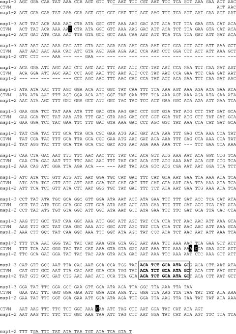
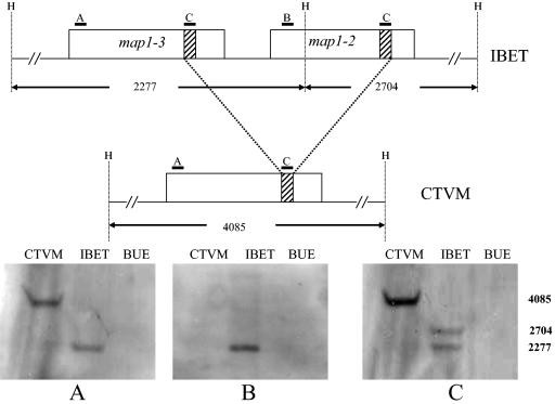
When testing the gene-specific primers, no product was detected for map1-2 when genomic DNA from the CTVM Gardel isolate was used. However, when genomic DNA from the IBET Gardel isolate was used the expected product was found. To investigate this further a new primer (map1-2R2) was designed in the intergenic region between map1-2 and map1-1. No product was amplified from CTVM Gardel when this primer was used in combination with primer map1-2F. However, a product was amplified when the primer was used in combination with primer map1-3F. The resulting product was approximately 1 kb, whereas the same primer combination yielded a product of approximately 1.8 kb (as expected from the determined sequence) from IBET Gardel (data not shown). Sequencing of the 1-kb PCR product showed that the first part (bp 1 to 713) of the sequence was 99.5% identical to map1-3 while the second part (bp 700 to 837) was identical to map1-2 and the intergenic region between map1-2 and map1-1 (with the exception of 1 nucleotide), suggesting that a recombination between map1-3 and map1-2 had occurred (Fig. 2). A sequence of 14 bp was detected which was present in both the sequences of IBET Gardel map1-2 and map1-3. The resulting “hybrid” gene had a continuous open reading frame (ORF) which was the same size as the map1-3 gene in IBET Gardel. To confirm that the observed deletion in the Gardel CTVM isolate was not due to artifacts introduced by the PCR, Southern blots using map1-2- and map1-3-specific probes and a probe specific for the recombination site were conducted. As expected, the map1-2-specific probe only reacted with a HindIII fragment (2,277 bp) in the Gardel IBET lane, the map1-3 specific-probe reacted with different HindIII fragments in the Gardel IBET (2,277 bp) and Gardel CTVM (4,085 bp) lanes, and the recombination site-specific probe reacted with two fragments (2,277 and 2,704 bp) in the Gardel IBET lane and only one fragment (4,085 bp) in the Gardel CTVM lane (Fig. 3). None of the probes reacted with HindIII-digested genomic DNA from uninfected endothelial cells. To confirm that both samples were indeed the Gardel isolate, the map1 genes from the two samples were amplified and sequenced. Both sequences were identical to the Gardel map1 sequence (accession number U50832) published in GenBank (data not shown).
FIG. 2.
Sequence alignment of the relevant part of E. ruminantium Gardel map1-3 (top), map1-2 (bottom), and the sequence obtained from CTVM Gardel with primers MAP1-3F and MAP1-2R2 (underlined). A black background indicates nucleotides that are different between the two sequences. The putative recombination site is boxed in and boldface.
FIG. 3.
Southern blot on HindIII-digested genomic DNA. The top shows a schematic representation of the genomic region containing map1-3 and map1-2 with the position of the HindIII restriction sites (H) and the location of the different probes (A to C). The bottom shows the genomic Southern blot with probe MAP1-3F (A), MAP1-2F (B), and the recombination site (C). BUE indicates uninfected endothelial cells.
Transcriptional analysis of the map1 gene cluster in bovine endothelial cell cultures.
To assess possible differences in transcription of members of the map1 gene cluster of different isolates during growth in endothelial cells, cDNA was prepared from endothelial cell cultures infected with the Gardel (IBET and CTVM), Senegal, and Welgevonden isolates of E. ruminantium and analyzed by PCR using map1 paralog- and in some cases isolate-specific primers. Independent of the source of endothelial cells (pulmonary artery or umbilical cord) or the passage number of the isolate, all map1 paralogs were transcribed in the three studied isolates.
Transcriptional analysis of the map1 gene cluster in tick cell cultures.
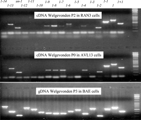
Gene transcription after growth of the CTVM Gardel or Welgevonden isolates in different tick cell lines was determined with map1 paralog-specific primers. For all tick cell lines tested, a transcript for map1-1 was present and was the predominant transcript (Table 3). Transcripts for map1-3 and map1-11 were never observed in any of the infected tick cell lines. Some transcripts were only detected in nonvector tick cell lines, namely map1-12, map1-10, map1-6, and map1-5 (Table 3). Figure 4 shows the results obtained from the AVL/CTVM13 and RAN/CTVM3 cell lines infected with the Welgevonden isolate of E. ruminantium and illustrates the variation in the amount of gene product amplified for the individual paralogs.
TABLE 3.
Transcription of the E. ruminantium map1 multigene family in infected tick cell lines
| Gene | Cell line, cell line passage no., isolate, and isolate passage no.a
|
|||||||||||
|---|---|---|---|---|---|---|---|---|---|---|---|---|
| AVL13
|
IDE8
|
IRE18
|
RAN3
|
RAE1
|
RAE25
|
BME2
|
BDE16
|
|||||
| 18 | 18 | 82 | 83 | 23 | 56 | 61 | 56 | 85 | 147 | 78 | 26 | |
| G | W | G | W | G | G | W | W | W | W | G | G | |
| 0 (5)b | 0 (2) | 5 | 6 | 5 | 14 (7) | 2 (1) | 5 (1) | 1 (1) | 1 (1) | 5 (4) | 8 (4) | |
| map1-14 | + | + | + | + | + | |||||||
| map1-13 | + | + | + | + | + | + | + | + | ||||
| un-1 | + | + | + | + | + | |||||||
| map1-12 | + | |||||||||||
| map1-11 | ||||||||||||
| map1-10 | + | + | + | |||||||||
| map1-9 | + | + | + | + | + | + | + | + | ||||
| map1-8 | + | + | + | + | + | + | + | |||||
| map1-7 | + | + | + | + | + | + | + | + | + | + | ||
| map1-6 | + | + | + | + | + | |||||||
| map1-5 | + | + | ||||||||||
| map1-4 | + | + | + | + | + | + | + | + | ||||
| map1-3 | ||||||||||||
| map1-2 | + | + | + | + | + | |||||||
| map1-1 | + | + | + | + | + | + | + | + | + | + | + | + |
| map1 | + | + | + | + | + | + | + | + | + | |||
| map1+1 | + | + | + | + | + | + | + | + | + | + | + | + |
Infected cell lines IDE8, RAN3, RAE1, RAE25, BME2, and BDE16 were incubated at 31°C, IRE18 at 28°C, and AVL13 at 37°C. “/CTVM” has been omitted from cell line names for simplicity. G, CTVM Gardel lacking map1-2; W, Welgevonden.
The first number indicates the E. ruminantium passage level in the cell line, the number in parentheses is the passage level in the IDE8 culture used to infect the cell line. IDE8 and IRE18 were infected directly from bovine endothelial cells (4).
FIG. 4.
RT-PCR of the map1 multigene family of E. ruminantium Welgevonden in the A. variegatum cell line AVL/CTVM13 (middle) and an R. appendiculatus cell line RAN/CTVM3 (top). The bottom panel shows a control PCR with the same primer set using genomic DNA (gDNA) as template. Lanes 1 to 17 contain PCR products for map1 paralogs as shown, lane 18 contains the PCR negative control, and lane 19 contains molecular weight markers.
DISCUSSION
We report here the observation that the E. ruminantium map1 gene cluster is relatively conserved among different isolates but can be subject to recombination leading to removal of entire genes from the cluster. Furthermore, the transcriptional analysis of the genes within the locus in vitro in host endothelial and vector and nonvector tick cell lines is reported. All genes were found to be transcribed in endothelial cell culture as described recently (38), independent of the source of the endothelial cells. In tick cell lines however, transcription of some paralogs could not be detected, and variation in the amount of amplified product was observed with map1-1 always being the most prominent transcript.
One of our most remarkable results was that two subpopulations of the Gardel isolate exhibited a different gene composition, despite the apparent conservation in gene organization of the map1 gene cluster among E. ruminantium isolates. The deletion of the map1-2 gene in the CTVM Gardel subpopulation indicates that recombination can occur which may influence the phenotype of the bacteria. At the site of the deletion a repeat motif was detected of 14 bp which occurred only in map1-2 and map1-3. The absence of the map1-2 gene in the CTVM Gardel map1 gene cluster can explain the inability to amplify this gene in previous experiments (2). The observation that the Southern blot with the map1-2-specific probe did not show any fragment in the Gardel CTVM lane suggests that the gene is not present elsewhere in the genome. Unfortunately it was not possible to completely trace the history of the CTVM Gardel isolate, but following arrival at CTVM the isolate was used to infect goats, and cultures were established from their blood. As the oldest identifiable cultures derived from these goats at CTVM already showed the absence of map1-2 by passage 2, we assume the recombination event occurred in the initial infections at CTVM or had already occurred in the material obtained from Guadeloupe.
Reddy et al. (29) described recombinase motifs and a high frequency of repeat elements in the p28 multigene locus of E. chaffeensis suggesting that recombination could occur but found no detectable rearrangements over a period of 2.5 years in an in vitro culture of the Arkansas isolate (29). Collins et al. (10) recently reported that the genome of E. ruminantium is very rich in tandemly repeated and duplicated sequences and that these repeats have mediated numerous translocation and inversion events that have resulted in the duplication and truncation of some genes and have also given rise to new genes (10). As the map1-2 gene is present in the Welgevonden and Senegal isolate and could also be amplified from other isolates (data not shown), we assume that the gene is deleted from CTVM Gardel. Cheng et al. (9) showed that molecular heterogeneity of E. chaffeensis isolates occurred in the α-repetitive region of the cluster, giving rise to three genetic groups containing either 22 (group I and III) or 21 genes (group II). Two locations were identified where insertions and/or deletions occurred (9). The recombination described in the present study appeared however in the β-repetitive region and was observed in what was considered to be a single isolate. As all E. ruminantium isolates for which the map1 gene sequence is known are unique, and we obtained identical map1 sequences from both Gardel sources, we assumed we were dealing with only one isolate. However, it was shown for another isolate (Kümm) of E. ruminantium that it in fact consisted of two genetically different “strains” which were separated and identified through their different host cell tropism in vitro (44), so it is possible that the original material received at CTVM contained two “strains,” of which one was subsequently established in culture and the other was not.
We found all the map1 paralogs and the gene of unknown function (un-1) to be transcriptionally active when the Senegal, Gardel, and Welgevonden isolates were cultured in endothelial cells, confirming the recent report of van Heerden et al. (38). We did not investigate whether or not paralogs were cotranscribed. Cultures were tested at different passage numbers and in different endothelial cell lines, but in all cases the results were the same. For unknown reasons transcription could not be detected in all cases when samples were tested the first time but were detected when samples were retested. These results differ from the data in endothelial cells previously reported by us, in which we only found map1 transcribed and no transcripts for map1-1 or map1-2 (2). This may be explained by the fact that in the present study we used two specific primers for each paralog and prepared cDNA using random hexamer primers, whereas previously we used map1 cluster-specific primers to generate the cDNA. Discrepancies in gene transcription have also been reported from studies in monocyte cultures of the p28 and p30 multigene families in E. chaffeensis and E. canis (23, 24, 30, 42). The same gene families were also studied in vivo in dogs and in tick vectors. In monocytes of infected dogs, 11 out of 14 E. canis p30 genes and 16 out of 22 E. chaffeensis p28 genes were transcribed, whereas transcription of only 1 gene (p30-10 or omp-1B) was detected in Rhipicephalus sanguineus or Amblyomma americanum ticks, respectively (13, 35, 36).
We found that the E. ruminantium ortholog of p30-10 and omp-1B, map1-1, was transcribed in all the E. ruminantium-infected tick cell lines and was the most dominant transcript present. However, we also found other map1 paralogs transcribed in tick cell lines indicating that, in vitro at least, several paralogs can be transcribed in tick cells. We previously reported that in A. variegatum ticks both map1-1 and map1 of the Senegal isolate are transcribed (2). In the vector cell line AVL/CTVM13 we found 10 out of 16 map1 paralogs transcribed for the Welgevonden isolate and 9 for the CTVM Gardel isolate, including map1-1 and map1. The transcription pattern for the two isolates was thus identical considering that map1-2 is missing from the CTVM Gardel isolate used in these cultures. Although Amblyomma species are the only known vectors for E. ruminantium (41), several cell lines established from other tick genera have been shown to support growth of E. ruminantium (4, 5, 7). The number of transcripts detected in these nonvector cell lines ranged from 4 (RAE25) to 11 (IDE8). For the Welgevonden isolate all the transcripts found in the vector cell line AVL/CTVM13 were also found in the nonvector line IDE8. Despite the differences observed in transcription patterns between the various tick cell lines, transcripts of two paralogs (map1-11 and map1-3) were never detected and two paralogs (map1-1 and map1+1) were always found to be transcribed. A surprising observation was the transcription of four paralogs (map1-12, map1-10, map1-6, and map 1-5) in nonvector tick cell lines which were not detected in the vector cell line AVL/CTVM13 but which were transcribed in endothelial cells. Studying the transcription of the map1 multigene family in infected ticks may show if the same paralogs are transcribed in vivo as in vitro in tick cell lines. At this point it should be noted that our RT-PCR results should be interpreted with caution, as finding a transcript does not necessarily imply that the mRNA is translated into a protein. In E. chaffeensis, despite the fact that multiple transcripts for p28 (omp-1) were detected, the product of only one gene was found based on N-terminal amino acid sequencing of expressed proteins from cultured organisms (23, 28). Using a proteomics approach, Singu et al. (31) showed for the same organism the expression of the products of two omp genes (p28-Omp19 and p28-Omp20) in macrophages and one (p28-Omp-14) in tick cells and that these proteins are posttranslationally modified by phosphorylation and glycosylation to generate multiple expressed forms (31). Whether a similar discrepancy between transcription and translation and/or expression exist for E. ruminantium remains to be determined.
As there have not been any studies reported on E. canis and E. chaffeensis gene transcription in vitro in tick cell lines, it is difficult to evaluate our results. However, some work has been done on two other rickettsial pathogens in I. scapularis cell lines, namely Anaplasma marginale and Anaplasma phagocytophilum. The A. marginale major surface protein 2 (MSP2) is encoded by a multigene family, and two MSP2 operon-associated proteins were expressed by A. marginale in IDE8 cells and in the bovine host during acute rickettsaemia, while only one was expressed in Dermacentor ticks during transmission feeding (22). Garcia-Garcia et al. (15) studied expression of A. marginale MSP1a, MSP1b, and MSP5 in infected bovine erythrocytes, IDE8 cells, and Dermacentor salivary glands. While the levels of MSP5 and MSP1b were similar in extracts of erythrocytes and IDE8 cells, MSP1a levels were much lower in IDE8 cells than in erythrocytes and were undetectable in sections of salivary glands. Transcriptional analysis indicated that the expression of MSP1a was probably regulated at the transcriptional level, since the amount of msp1α transcripts in infected IDE8 cells was lower than that in erythrocytes and undetectable in infected salivary glands, while transcripts for msp1β, msp4, and 16S rRNA were present in all three tissues (15). The immunodominant surface protein P44 of A. phagocytophilum is also encoded by a multigene family and is expressed in vitro in the human promyelocytic cell line HL-60. When the pathogen was transferred to the I. scapularis cell line ISE6 growing at 34°C, downregulation of a specific P44 antigen was observed in Western blots using a monoclonal antibody. This effect was not temperature dependent, since it was also seen in tick cells maintained at 37°C, but upregulation occurred after the pathogen was returned to HL-60 cells (16). Later it was shown that the p44 gene in the expression site is polymorphic in vitro (HL-60 cells and ISE6 tick cells [1]) and in vivo (mice and I. scapularis ticks [14] and humans [1, 21]). Temperature does not seem to play a major role in the transcription of the E. ruminantium map1 multigene family in vitro either, as the same paralogs transcribed at 37°C in AVL/CTVM13 cells were also detected at 31°C in IDE8 cells, and no differences were seen in transcription pattern between incubation temperatures of 30°C and 37°C for E. ruminantium in endothelial cells (2, 38).
Together, these data indicate that the E. ruminantium map1 multigene family is regulated differently in the host and vector cell environments in vitro and can be subject to recombination leading to an altered gene arrangement. Studying the transcriptional activity using quantitative assays of the E. ruminantium map1 multigene family in its natural environment in the host and vector in vivo combined with a proteomics approach should provide more insight into the regulation and function of this gene family.
Acknowledgments
The research described in the manuscript was supported by a European Union INCO DEV grant (ICA4-CT-2000-30026) and DfID Animal Health Programme project R7363 and CIRAD-IGH genome project no. 751745/00.
We are grateful to Paula Alves for providing BAE cells and the IBET Gardel isolate of E. ruminantium and to Ulrike Munderloh and Tim Kurtti for providing the RAE25 and IDE8 cell lines. Henriette van Heerden is thanked for providing the sequences of the Welgevonden map1 cluster primers ahead of publication, and Jos van Putten is thanked for critical reading of the manuscript and helpful comments. We thank Jacques Demaille from Institute of Human Genetics (IGH) and Gérard Matheron (Agropolis) for having supported the full sequencing of the E. ruminantium (Gardel) genome.
REFERENCES
- 1.Barbet, A. F., P. F. M. Meeus, M. Bélanger, M. V. Bowie, J. Yi, A. M. Lundgren, A. R. Alleman, S. J. Wong, F. K. Chu, U. G. Munderloh, and S. D. Jauron. 2003. Expression of multiple outer membrane protein sequence variants from a single genomic locus of Anaplasma phagocytophilum. Infect. Immun. 71:1706-1718. [DOI] [PMC free article] [PubMed] [Google Scholar]
- 2.Bekker, C. P., L. Bell-Sakyi, E. A. Paxton, D. Martinez, A. Bensaid, and F. Jongejan. 2002. Transcriptional analysis of the major antigenic protein 1 multigene family of Cowdria ruminantium. Gene 285:193-201. [DOI] [PubMed] [Google Scholar]
- 3.Bell, L. J. 1983. Development of Theileria in tick tissue culture. MPhil. thesis. University of Edinburgh, United Kingdom.
- 4.Bell-Sakyi, L. 2004. Ehrlichia ruminantium grows in cell lines from four ixodid tick genera. J. Comp. Pathol. 130:285-293. [DOI] [PubMed] [Google Scholar]
- 5.Bell-Sakyi, L., E. A. Paxton, U. G. Munderloh, and K. J. Sumption. 2000. Growth of Cowdria ruminantium, the causative agent of heartwater, in a tick cell line. J. Clin. Microbiol. 38:1238-1240. [DOI] [PMC free article] [PubMed] [Google Scholar]
- 6.Bell-Sakyi, L., E. A. Paxton, U. G. Munderloh, and K. J. Sumption. 2000. Presented at the ticks and tick-borne pathogens: into the 21st century. Institute of Zoology, Slovak Academy of Science, Bratislava, Slovakia.
- 7.Bell-Sakyi, L., E. A. Paxton, P. Wright, and K. J. Sumption. 2002. Immunogenicity of Ehrlichia ruminantium grown in tick cell lines. Exp. Appl. Acarol. 28:177-185. [DOI] [PubMed] [Google Scholar]
- 8.Bezuidenhout, J. D. 1987. Natural transmission of heartwater. Onderstepoort J. Vet. Res. 54:349-351. [PubMed] [Google Scholar]
- 9.Cheng, C., C. D. Paddock, and R. Reddy Ganta. 2003. Molecular heterogeneity of Ehrlichia chaffeensis isolates determined by sequence analysis of the 28-kilodalton outer membrane protein genes and other regions of the genome. Infect. Immun. 71:187-195. [DOI] [PMC free article] [PubMed] [Google Scholar]
- 10.Collins, N. E., J. Liebenberg, E. P. de Villiers, K. A. Brayton, E. Louw, A. Pretorius, F. E. Faber, H. van Heerden, A. Josemans, M. van Kleef, H. C. Steyn, M. F. van Strijp, E. Zweygarth, F. Jongejan, J. C. Maillard, D. Berthier, M. Botha, F. Joubert, C. H. Corton, N. R. Thomson, M. T. Allsopp, and B. A. Allsopp. 2005. The genome of the heartwater agent Ehrlichia ruminantium contains multiple tandem repeats of actively variable copy number. Proc. Natl. Acad. Sci. USA 102:838-843. [DOI] [PMC free article] [PubMed] [Google Scholar]
- 11.Du Plessis, J. L. 1985. A method for determining the Cowdria ruminantium infection rate of Amblyomma hebraeum: effects in mice injected with tick homogenates. Onderstepoort J. Vet. Res. 52:55-61. [PubMed] [Google Scholar]
- 12.Dumler, J. S., A. F. Barbet, C. P. J. Bekker, G. A. Dasch, G. H. Palmer, S. C. Ray, Y. Rikihisa, and F. R. Rurangirwa. 2001. Reorganization of genera in the families Rickettsiaceae and Anaplasmataceae in the order Rickettsiales: unification of some species of Ehrlichia with Anaplasma, Cowdria with Ehrlichia and Ehrlichia with Neorickettsia, descriptions of six new species combinations and designation of Ehrlichia equi and ‘HGE agent’ as subjective synonyms of Ehrlichia phagocytophila. Int. J. Syst. Evol. Microbiol. 51:2145-2165. [DOI] [PubMed] [Google Scholar]
- 13.Felek, S., R. Greene, and Y. Rikihisa. 2003. Transcriptional analysis of p30 major outer membrane protein genes of Ehrlichia canis in naturally infected ticks and sequence analysis of p30-10 of E. canis from diverse geographic regions. J. Clin. Microbiol. 41:886-888. [DOI] [PMC free article] [PubMed] [Google Scholar]
- 14.Felek, S., S. Telford III, R. C. Falco, and Y. Rikihisa. 2004. Sequence analysis of p44 homologs expressed by Anaplasma phagocytophilum in infected ticks feeding on naive hosts and mice infected by tick attachment. Infect. Immun. 72:659-666. [DOI] [PMC free article] [PubMed] [Google Scholar]
- 15.Garcia-Garcia, J. C., J. de la Fuente, E. F. Blouin, T. J. Johnson, T. Halbur, V. C. Onet, J. T. Saliki, and K. M. Kocan. 2004. Differential expression of the msp1α gene of Anaplasma marginale occurs in bovine erythrocytes and tick cells. Vet. Microbiol. 98:261-272. [DOI] [PubMed] [Google Scholar]
- 16.Jauron, S. D., C. M. Nelson, V. Fingerle, M. D. Ravyn, J. L. Goodman, R. C. Johnson, R. Lobentanzer, B. Wilske, and U. G. Munderloh. 2001. Host cell-specific expression of a p44 epitope by the human granulocytic ehrlichiosis agent. J. Infect. Dis. 184:1445-1450. [DOI] [PubMed] [Google Scholar]
- 17.Jongejan, F. 1991. Protective immunity to heartwater (Cowdria ruminantium infection) is acquired after vaccination with in vitro-attenuated rickettsiae. Infect. Immun. 59:729-731. [DOI] [PMC free article] [PubMed] [Google Scholar]
- 18.Jongejan, F., G. Uilenberg, F. F. Franssen, A. Gueye, and J. Nieuwenhuijs. 1988. Antigenic differences between stocks of Cowdria ruminantium. Res. Vet. Sci. 44:186-189. [PubMed] [Google Scholar]
- 19.Kocan, K. M., J. D. Bezuidenhout, and A. Hart. 1987. Ultrastructural features of Cowdria ruminantium in midgut epithelial cells and salivary glands of nymphal Amblyomma hebraeum. Onderstepoort J. Vet. Res. 54:87-92. [PubMed] [Google Scholar]
- 20.Kurtti, T. J., U. G. Munderloh, and M. Samish. 1982. Effect of medium supplements on tick cells in culture. J. Parasitol. 68:930-935. [PubMed] [Google Scholar]
- 21.Lin, Q., Y. Rikihisa, N. Ohashi, and N. Zhi. 2003. Mechanisms of variant p44 expression by Anaplasma phagocytophilum. Infect. Immun. 71:5650-5661. [DOI] [PMC free article] [PubMed] [Google Scholar]
- 22.Löhr, C. V., K. A. Brayton, V. Shkap, T. Molad, A. F. Barbet, W. C. Brown, and G. H. Palmer. 2002. Expression of Anaplasma marginale major surface protein 2 operon-associated proteins during mammalian and arthropod infection. Infect. Immun. 70:6005-6012. [DOI] [PMC free article] [PubMed] [Google Scholar]
- 23.Long, S. W., X. F. Zhang, H. Qi, S. Standaert, D. H. Walker, and X. J. Yu. 2002. Antigenic variation of Ehrlichia chaffeensis resulting from differential expression of the 28-kilodalton protein gene family. Infect. Immun. 70:1824-1831. [DOI] [PMC free article] [PubMed] [Google Scholar]
- 24.McBride, J. W., X. J. Yu, and D. H. Walker. 2000. A conserved, transcriptionally active p28 multigene locus of Ehrlichia canis. Gene 254:245-252. [DOI] [PubMed] [Google Scholar]
- 25.Munderloh, U. G., Y. Liu, M. Wang, C. Chen, and T. J. Kurtti. 1994. Establishment, maintenance and description of cell lines from the tick Ixodes scapularis. J. Parasitol. 80:533-543. [PubMed] [Google Scholar]
- 26.Mutunga, M., P. M. Preston, and K. J. Sumption. 1998. Nitric oxide is produced by Cowdria ruminantium-infected bovine pulmonary endothelial cells in vitro and is stimulated by gamma interferon. Infect. Immun. 66:2115-2121. [DOI] [PMC free article] [PubMed] [Google Scholar]
- 27.Ohashi, N., Y. Rikihisa, and A. Unver. 2001. Analysis of transcriptionally active gene clusters of major outer membrane protein multigene family in Ehrlichia canis and E. chaffeensis. Infect. Immun. 69:2083-2091. [DOI] [PMC free article] [PubMed] [Google Scholar]
- 28.Ohashi, N., N. Zhi, Y. Zhang, and Y. Rikihisa. 1998. Immunodominant major outer membrane proteins of Ehrlichia chaffeensis are encoded by a polymorphic multigene family. Infect. Immun. 66:132-139. [DOI] [PMC free article] [PubMed] [Google Scholar]
- 29.Reddy, G. R., and C. P. Streck. 1999. Variability in the 28-kDa surface antigen protein multigene locus of isolates of the emerging disease agent Ehrlichia chaffeensis suggests that it plays a role in immune evasion. Mol. Cell. Biol. Res. Commun. 1:167-175. [DOI] [PubMed] [Google Scholar]
- 30.Reddy, G. R., C. R. Sulsona, A. F. Barbet, S. M. Mahan, M. J. Burridge, and A. R. Alleman. 1998. Molecular characterization of a 28 kDa surface antigen gene family of the tribe Ehrlichiae. Biochem. Biophys. Res. Commun. 247:636-643. [DOI] [PubMed] [Google Scholar]
- 31.Singu, V., H. Liu, C. Cheng, and R. R. Ganta. 2005. Ehrlichia chaffeensis expresses macrophage- and tick cell-specific 28-kilodalton outer membrane proteins. Infect. Immun. 73:79-87. [DOI] [PMC free article] [PubMed] [Google Scholar]
- 32.Sulsona, C. R., S. M. Mahan, and A. F. Barbet. 1999. The map1 gene of Cowdria ruminantium is a member of a multigene family containing both conserved and variable genes. Biochem. Biophys. Res. Commun. 257:300-305. [DOI] [PubMed] [Google Scholar]
- 33.Uilenberg, G. 1983. Heartwater (Cowdria ruminantium infection): current status. Adv. Vet. Sci. Comp. Med. 27:428-455. [PubMed] [Google Scholar]
- 34.Uilenberg, G., E. Camus, and N. Barre. 1985. Quelques observations sur une souche de Cowdria ruminantium isolée en Guadeloupe (Antilles françaises). Rév. Elev. Med. Vet. Pays Trop. 38:34-42. [PubMed] [Google Scholar]
- 35.Unver, A., N. Ohashi, T. Tajima, R. W. Stich, D. Grover, and Y. Rikihisa. 2001. Transcriptional analysis of p30 major outer membrane multigene family of Ehrlichia canis in dogs, ticks, and cell culture at different temperatures. Infect. Immun. 69:6172-6178. [DOI] [PMC free article] [PubMed] [Google Scholar]
- 36.Unver, A., Y. Rikihisa, R. W. Stich, N. Ohashi, and S. Felek. 2002. The omp-1 major outer membrane multigene family of Ehrlichia chaffeensis is differentially expressed in canine and tick hosts. Infect. Immun. 70:4701-4704. [DOI] [PMC free article] [PubMed] [Google Scholar]
- 37.Van Heerden, H., N. E. Collins, M. T. Allsopp, and B. A. Allsopp. 2002. Major outer membrane proteins of Ehrlichia ruminantium encoded by a multigene family. Ann. N. Y. Acad. Sci. 969:131-134. [DOI] [PubMed] [Google Scholar]
- 38.Van Heerden, H., N. E. Collins, K. A. Brayton, C. Rademeyer, and B. A. Allsopp. 2004. Characterization of a major outer membrane protein multigene family in Ehrlichia ruminantium. Gene 330:159-168. [DOI] [PubMed] [Google Scholar]
- 39.van Vliet, A. H. M., F. Jongejan, and B. A. M. van der Zeijst. 1992. Phylogenetic position of Cowdria ruminantium (Rickettsiales) determined by analysis of amplified 16S ribosomal DNA sequences. Int. J. Syst. Bacteriol. 42:494-498. [DOI] [PubMed] [Google Scholar]
- 40.van Vliet, A. H. M., F. Jongejan, M. van Kleef, and B. A. M. van der Zeijst. 1994. Molecular cloning, sequence analysis, and expression of the gene encoding the immunodominant 32-kilodalton protein of Cowdria ruminantium. Infect. Immun. 62:1451-1456. [DOI] [PMC free article] [PubMed] [Google Scholar]
- 41.Walker, J. B., and A. Olwage. 1987. The tick vectors of Cowdria ruminantium (Ixodoidea, Ixodidae, genus Amblyomma) and their distribution. Onderstepoort J. Vet. Res. 54:353-379. [PubMed] [Google Scholar]
- 42.Yu, X., J. W. McBride, X. Zhang, and D. H. Walker. 2000. Characterization of the complete transcriptionally active Ehrlichia chaffeensis 28 kDa outer membrane protein multigene family. Gene 248:59-68. [DOI] [PubMed] [Google Scholar]
- 43.Yu, X. J., J. W. McBride, and D. H. Walker. 1999. Genetic diversity of the 28-kilodalton outer membrane protein gene in human isolates of Ehrlichia chaffeensis. J. Clin. Microbiol. 37:1137-1143. [DOI] [PMC free article] [PubMed] [Google Scholar]
- 44.Zweygarth, E., A. I. Josemans, H. van Heerden, M. T. E. P. Allsopp, and B. A. Allsopp. 2002. The Kümm isolate of Ehrlichia ruminantium: in vitro isolation, propagation and characterization. Onderstepoort J. Vet. Res. 69:147-153. [PubMed] [Google Scholar]


