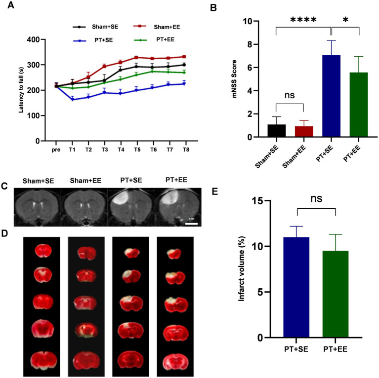
Figure 2.
EE protects against brain damage after an ischemic stroke. (A) The time of latency to fall in the rota-rod test during the 2-day training (n = 18). (B) The brain damage was evaluated by mNSS scores (n = 12). (C) The T2-weighted images of whole-brain MRI on day 3 after the PT stroke showed the infarct area (increased signal intensity). (D) 2,3,5-Triphenyltetrazolium chloride (TTC) staining of brain slices 3 days after PT stroke showed the infarct area (white). (E) The relative proportion and quantification of infarct volume by TTC staining (n = 3). ns: no significant, *p < 0.05, ***p < 0.001, ****p < 0.0001. Error bars were represented as mean ± SD.

