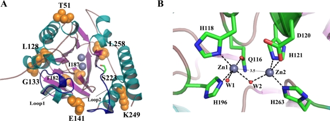
Fig. 6.
Overall and active-site structure of GOB-38. (A) The GOB-38 protein was depicted in a cartoon representation, where the helices were colored in dark green, the strands in pink, and the non-structured loops in dark yellow. Specifically, the two loops, referred to as loop1 and loop2, which covered the active site, were highlighted in dark blue. The presence of zinc atoms was represented by gray spheres. The residues that were the subject of discussion in this paper were visually represented using orange spheres and stick models, with carbon atoms depicted in green, nitrogen atoms in blue, and oxygen atoms in red. (B) The active-site structure of GOB-38 was depicted, with gray representing the zinc atoms, red representing the water molecules (Wat1, Wat2), and dashed lines indicating the coordination bonds. The estimated distance between the two Zn2+ ions was 3.5 Å.

