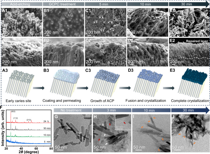
Fig. 3. Rapid repair of demineralized enamel by GCPC in artificial saliva.
A1–A3, B1–B3, C1–C3, D1–D3, E1–E3 SEM images from the top surface and cross-sectional view of etched enamel before (A1–A2) and after treatment for 0 min (B1–B2), 5 min (C1–C2), 10 min (D1–D2) and 30 min (E1–E2). The dashed line in E2 marks the speculated boundary between the natural enamel and the repair layer. A3, B3, C3, D3 Schemes illustrating the repair performances of GCPC on etched enamels over time, which goes through enamel caries site caused by acid etching (A3), the coating and permeating in 0 min (B3), growth of ACP in 5 min (C3), fusion and crystallization in 10 min (D3) and complete crystallization in 30 min (E3). F GIXRD spectra of enamel surface repaired by GCPC for 5 min, 10 min, 30 min and 24 h. G TEM image of the HAP nanorods without treatment. Cryo-TEM images of the GCPC-treated HAP nanorods at 3 min (H) and 10 min (I). Clusters are indicated by white arrows and the newly formed nanowires are marked by red arrows in (H). J TEM images of the GCPC-treated HAP nanorods at 30 min. Newly formed structures at the ends of HAP nanorods are marked by orange arrows in (I, J).

