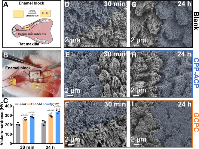
Fig. 7. The repair of demineralized enamel in vivo.
Scheme (A) and photograph (B) of vivo animal model showing the etched enamel fixed in the oral cavity of rats. C Microhardness of the enamels repaired by different materials in vivo for 30 min and 24 h. The error bars represent the mean ± SD for n = 3 independent experiments, and no data were excluded from the analyses. Statistical differences are assessed using one-way ANOVA and Tukey’s multiple comparisons test. The asterisk (*) denotes significant differences between the indicated group and the blank group (p values higher than 0.001 are displayed above the asterisks). **p < 0.01, ***p < 0.001. SEM images of the enamels repaired by deionized water (D), CPP-ACP paste (E) and GCPC (F) for 30 min in vivo. SEM images of the enamels repaired by deionized water (G), CPP-ACP paste (H) and GCPC (I) for 24 h in vivo. The blue regions represent the treatment areas.

