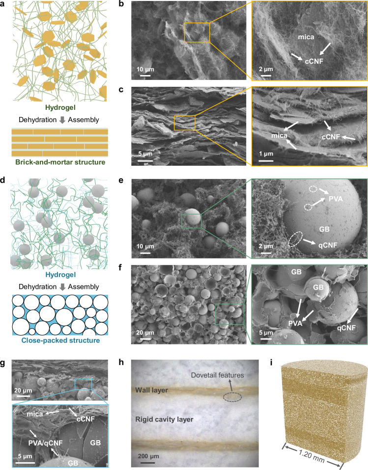
Fig. 2. Synchronous assembly of various microstructures in the RCWSM.
a Schematic of the brick-and-mortar wall structure from the initial hydrogel to the dense state. Amino mica, yellow platelet; carboxylated CNF (cCNF), green curve. b SEM images of the irregular arrangement of mica platelets in the initial hydrogel. c SEM images of the brick-and-mortar wall in the RCWSM. d Schematic of the rigid cavity layer from the initial hydrogel to the dense state. GB gray sphere, PVA blue curve, qCNF green curve. e SEM images of the irregular arrangement of GB in the initial hydrogel. f SEM images of the rigid cavity layer within the RCWSM. g SEM images of the interface between the brick-and-mortar wall and the close-packed rigid cavity layer. h Polarizing microscope image of the cross-section of the RCWSM, which shows the obvious dovetail features at the interface between the two layers. i 3D reconstruction of the RCWSM derived from X-ray microtomography.

