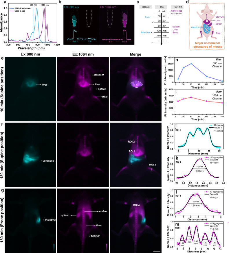
Fig. 5. The in vivo dual-channel fluorescence (Fl.) imaging of BALB/c mouse using ESi5-S agg.
a The normalized absorption spectra of ESi5-S monomer and ESi5-S agg and the compatible common laser lines. b The epi-fluorescence images of the ESi5-S Monomer (Left, 500 μM in water with 1% tween 80) and ESi5-S agg (500 μM in PBS) solutions in EP tubes excited by 808 (60 mW•cm−2) and 1064 nm (80 mW•cm−2), respectively. c The timeline of the in vivo fluorescence mouse imaging experiment. 808 nm channel: λex = 808 nm (60 mW•cm−2), long pass filter: 1200 nm, exposure time: 100 ms. 1064 nm channel: λex = 1064 nm (80 mW•cm−2), long pass filter: 1200 + 1350 nm, exposure time: 500 ms. d A schematic diagram of major anatomical structures of mouse related to this experiment. Created in BioRender. Yang, Y. (2024) https://BioRender.com/q64i385. The two-channel fluorescence images of BALB/c mice in the supine position (e) 10 min and (f) 180 min post i.v. injection, and in the prone position (g) at 180 min post i.v. injection. The changes of the liver region fluorescence intensity (h) in the 808 nm channel and (i) in the 1064 nm channel. The line intensities of (j) the ROI 1 for the intestine in the 808 nm channel, (k) the ROI 2 for the sternum in the 1064 nm channel, (l) the ROI 3 for the tibia in the 1064 nm channel, and (m) the ROI 4 for the lumbar vertebra in the 1064 nm channel. Scale bar: 10 mm.

