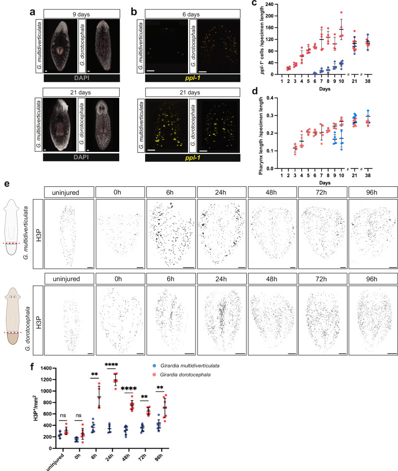
Fig. 9. Slower regeneration in cave planarians.
a G. multidiverticulata takes longer to regenerate its pharynx (visualized using DAPI); however, after 21 days, structures reach similar sizes. Scale bars, 50 µm. b G. multidiverticulata takes longer to regenerate its brain (marked with ppl-1); however, after 21 days both structures reach similar sizes. Scale bars, 50 µm. c Quantification (mean ± SD) of pharynx length (DAPI) normalized by specimen length, during regeneration in G. dorotocephala and G. multidiverticulata. d Quantification (mean ± SD) of brain cells (ppl-1+ normalized by specimen length), during regeneration of G. dorotocephala and G. multidiverticulata. The sample size for each experiment is provided in the “Quantification, statistics, and reproducibility” section. e Cave planarians failed to show robust elevation of mitosis following injury. Immunolabeling of mitotic cells (H3P+) in G. multidiverticulata non-discernible eye morphotype and in G. dorotocephala. Scale bars, 100 µm. f G. multidiverticulata exhibits lower numbers of mitotic cells (H3P+) per mm2, exclusively during regeneration, when compared with G. dorotocephala. H3P+ cells from each time point were counted, and compared between the two species using a Student’s two-tailed t-test; ns, not significant,**p < 0.01, ***p < 0.001, ****p < 0.0001. Mean ± SD. The G. dorotocephala counts involved n = 5, n = 7, n = 5, n = 6, n = 8, n = 6, and n = 8 animals for uninjured, 0 h, 6 h, 24 h, 48 h, 72 h, and 96 h, respectively. The G. dorotocephala counts involved n = 4, n = 6, n = 6, n = 5, n = 7, n = 7, and n = 7 animals for uninjured, 0 h, 6 h, 24 h, 48 h, 72 h, and 96 h, respectively.

