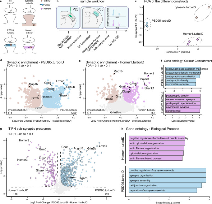
Fig. 2. Spatially accurate identification of L5 cortical neuron postsynaptic proteins.
a Representation of the different conditions tested and the expected pattern of biotinylated proteins at the synapse. b Mice are injected with AAVs in the somatosensory cortex at P28. At P35, biotin is provided 3 h before collection of somatodendritic material from somatosensory cortices. Biotinylated proteins are enriched by streptavidin pulldown and prepared for MS analysis. c PCA analysis shows replicates clustering by TurboID construct. d Relative protein enrichment for PSD95.turboID and cytosolic.turboID. Significant proteins (two-sided, unpaired t-test with permutation-based FDR correction at 10%, s0 > 0.1) are labelled, core excitatory postsynaptic proteins are highlighted. e Relative protein enrichment for Homer1.turboID and cytosolic.turboID. Significant proteins (two-sided, unpaired t-test with permutation-based FDR correction at 10%, s0 > 0.1) are labelled, core excitatory postsynaptic proteins are highlighted. f Gene ontology analysis of synaptic proteins in PSD95.turboID and Homer1.turboID samples. Gene ontology analysis was performed using Fisher’s one-tailed test with FDR multiple-comparison correction. g Relative protein enrichment by PSD95.turboID and Homer1.turboID. Significant proteins (two-sided, unpaired t-test with permutation-based FDR correction at 5%, s0 > 0.1) are labelled based on TurboID construct. h Gene ontology of differentially expressed proteins in PSD95.turboID vs. Homer1.turboID samples. Gene ontology analysis was performed using Fisher’s one-tailed test with FDR multiple-comparison correction. PCA principal component analysis, IT intratelencephalic, PN pyramidal neuron.

