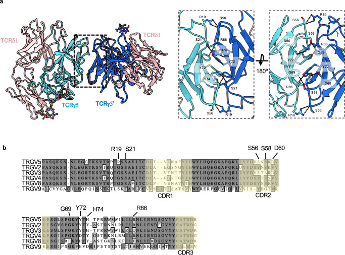
Fig. 4. Vγ5 Vδ1 TCR dimer interface and its sequence conservation.
a Structure of the 9C2 TCR ECD (Fab 2 complex used, Fab models hidden for clarity) with each TCRγ protomer colored a different shade of blue. Analysis of the TCRVγ5-mediated dimer interface with interacting residues shown as sticks and dashed lines representing putative hydrogen bonds. Note: TCRγ Y72 of protomer 1 forms a π-π interaction with Y72 of protomer 2. b Sequence alignment of various germline encoded TCR γ-chain variable regions. Residues that have been identified in the dimerization interface are labeled. CDRs are indicated with a faint yellow tint.

