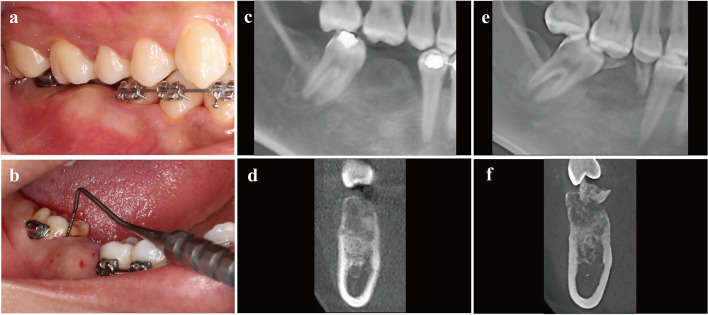
Fig. 1.
The clinical and radiographic views of the bone expansion in alveolar crest of edentulous site. a The buccal view presented with inadequate inter-arch space, which was caused by lesion bone extend, then the keratinized mucosa was normal. b The pseudo-pocket of the adjacent tooth and bleeding on probing of #47. c, d The radiographic views at initial diagnosis with the ground-glass bone of blurred boundaries. e, f The retrospective radiographic record of preoperative tooth extraction in 2015 with the residual root of #46 that existed for many years

